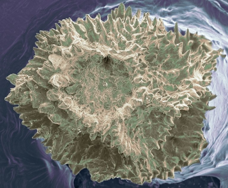
Sometimes it just takes a little technology to help us see familiar objects from a brand new perspective. The Cauliflower Coral (Pocillopora sp.) is no stranger to reefkeepers/SPS lovers, but it’s a safe bet few of us have viewed this coral under extreme magnification.
The content below is provided by Eric W. Roth (Mr. Microscope at Nano-reef.com) from his thread: Scanning Electron Microscopy of Pocillopora. Acknowledgements to Northwestern University NUANCE Center for the use of their Electron Microscope.
Eric plans to explore more reef life under the electron microscope. Stay tuned for more spectacular images.
As some of you know I work with microscopes. Well, the other day I got an opportunity to throw a piece of coral into one of our scanning electron microscopes. This is a small piece of Pocillopora that had bleached in my 3 gallon pico. I colorized a few images with Photoshop. Enjoy!
Getting in a little closer at an edge

Here we are zoomed in considerably further. There’s a bunch of salt crystals here. They seemed to come in two sizes.

Here are some of the larger salt crystals. If you look closely, you can see some of the smaller salt crystals too. They look like little white spots.

If you look in the lower right had corner of the above image, you’ll see a tiny diatom. I zoomed in on it here.

Here’s another diatom I found.

Here are the super tiny salt crystals. You can also start to see the Calcium Carbonate crystal growth of the coral skeleton here. Some of the CaCO3 makes hexagonal patterns.

Wrapping things up with a little perspective.


0 Comments