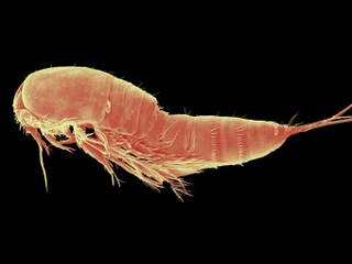
The content below is provided by Eric W. Roth (Mr. Microscope at Nano-reef.com) from his thread: Scanning Electron Microscopy of Pocillopora. Acknowledgements to Northwestern University NUANCE Center for the use of their Electron Microscope.
View Eric Roth’s other electron microscopy.
Scanning Electron Microscopy of Zooplankton
Hello All!
It’s taken me a while to gather all of this data; a minute here and there after sessions of looking at actual research samples, but I’ve finally managed to gather a decent collection of images. This entry’s samples consist of some pods that I managed to get out of my fuge. I found an infant amphipod, a copepod, something that looks vaguely like a caterpillar, and something that may be a microscopic bivalve of sorts. Enough chatter, here’s the good stuff!
Amphipod
First off, here is the amphipod I found. I used some static to get him to stand on his hind legs so that I could look him straight in the eye.

Roooar!!

Actually, I think he looks a little like Tony the Tiger. What do you think?

Let’s zoom in on that mouth a little.

“The better to EAT you with!!!”

Arms:

The Claw!

Copepod
Finally, here is the copepod. This little pod was a lot of fun to explore. I understand how these things can stick to our glass walls now. Their spikes have spikes of their own. I even think their spike’s spikes have spikes. There’s something to think about. I took the time at home to colorize this one.

Did you know that copepods only have one eye? You can see it in the front.
Spikes!

Here’s a closer look at the tail:

There was some bacteria caught up here:

This reminds me of a hunting knife:


0 Comments