So, you have shelled out a week’s pay for a really nice ‘chalice coral’ (Echinophyllia spp.) and you’re wondering if it will keep its beautiful colors. This article can assist you in your quest and help you understand how lighting and water’s physical qualities can affect your new coral.
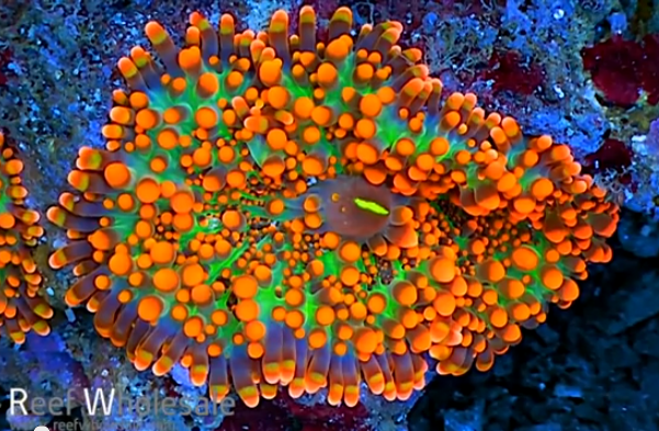
This time, we’ll examine those fluorescent proteins found mostly in those stony corals of suborder Faviina (although a number are from Families Lobophyllidae, Euphylliidae, and Agarciidae), with a few being isolated from soft corals and false corals. These stony genera include some of the highly colorful stony corals such as Echinophyllia, Trachyphyllia, Lobophyllia, Favia, Favites, Montastrea, Scolymia, and others. Soft corals include Dendronephthya, Clavularia, and Sarcophyton. The ‘false coral’ Ricordea are also known to contain this type of fluorescent protein. Altogether, almost 80 coral genera (at least 9 Families) are represented. Research suggests there are no nonfluorescent pigments (chromoproteins) in this group.
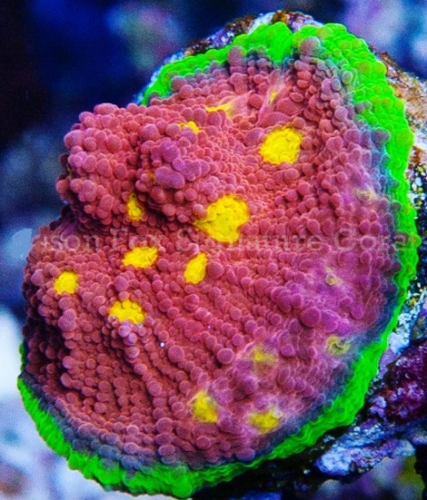
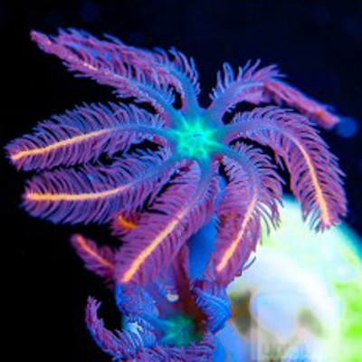
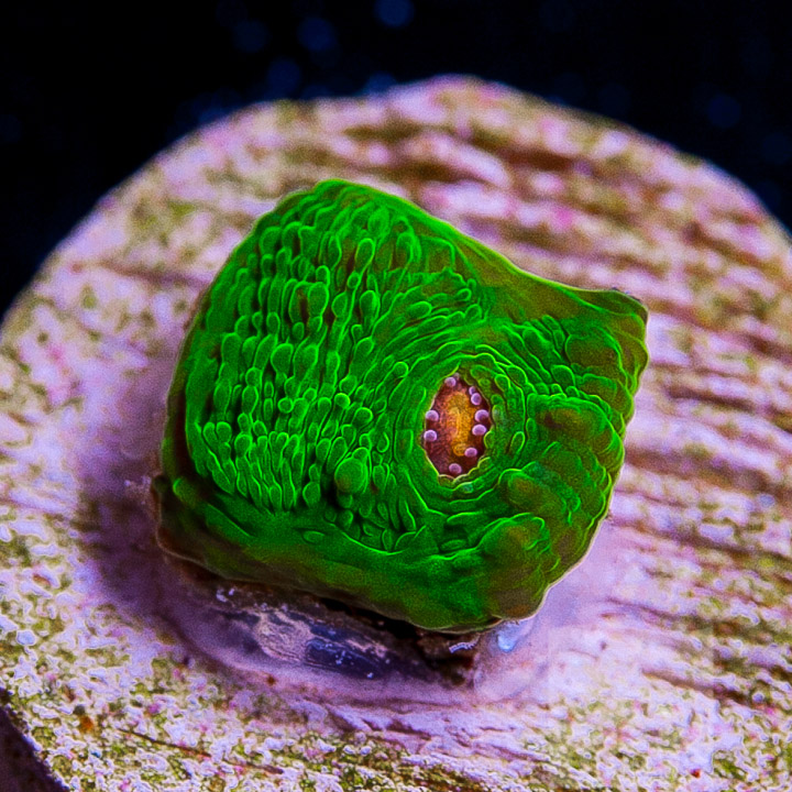
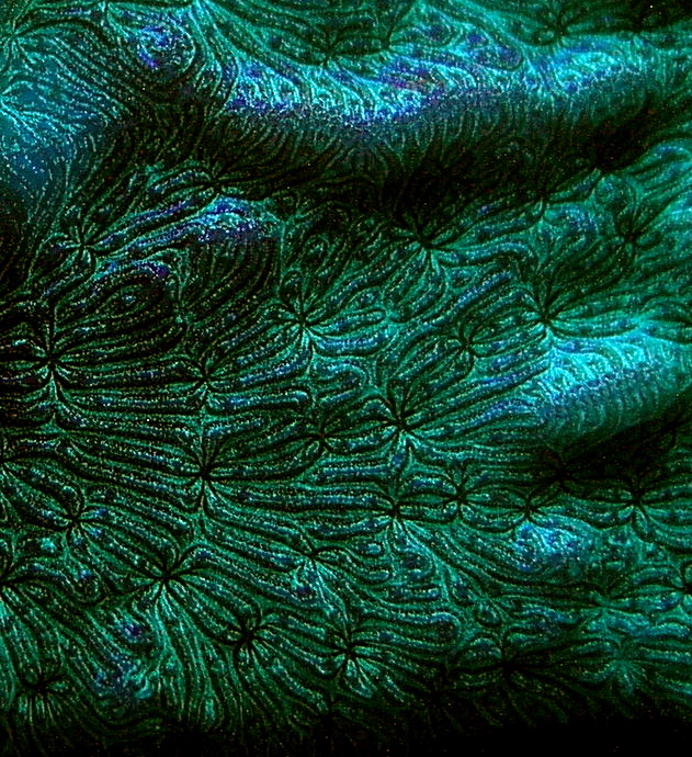
An example of Clade D fluorescent proteins. Photo courtesy of Jason Fox Signature Corals (jasonfoxsignaturecorals.com).
In this article, we’ll compare information from various sources and make educated decisions on how to keep your Clade D corals colorful. I’ll use the term ‘pigment’ from time to time – this is not technically correct, so please allow me some latitude.
Before beginning, perhaps a review of important terms is in order.
Glossary
- Absorption:
- The process in which incident radiation is retained without reflection or transmission.
- Clade:
- For our purposes in this article, a grouping of pigments based on similar features inherited from a common ancestor. Pigments from corals includes Clades A, B, C, and D. Clades can refer to living organisms as well (clades of Symbiodinium – zooxanthellae – are a good example.)
- Chromophore:
- The colorful portion of a pigment molecule. In some cases, chromophore refers to a granular packet containing many pigment molecules.
- Chromoprotein pigment:
- A non-fluorescent but colorful pigment. These pigments appear colorful because they reflect light. For example, a chromoprotein with a maximum absorption at 580nm might appear purple because it preferentially reflects blue and red wavelengths.
- Chromo-Red Pigment:
- A newly described type of pigment possessing characteristics of both chromoproteins and Ds-Red fluorescent proteins. Peak fluorescence is at 609nm (super red).
- Cyan Fluorescent Protein (CFP):
- Blue-green pigments with fluorescent emissions in the range of ~477-500nm. Cyan and green pigments share a similar chromophore structure. Cyan pigments are expressed at lower light levels than green, red or non-fluorescent pigments.
- Emission:
- That light which is fluoresced by a fluorescent pigment.
- Excitation:
- That light absorbed by a fluorescent pigment. Some of the excitation light is fluoresced or emitted at a less energetic wavelength (color).
- Fluorescence:
- Absorption of radiation at one wavelength (or color) and emission at another wavelength (color). Absorption is also called excitation. Fluorescence ends very soon after the excitation source is removed (on the order of ~2-3 nanoseconds: Salih and Cox, 2006).
- Green Fluorescent Protein (GFP):
- Fluorescent pigments with emissions of 500-525nm.
- ‘Hula Twist’:
- A bending of a fluorescent protein resulting in a change of apparent color. Molecular bonds are not broken; therefore the pigment can shift back and forth, with movements reminiscent of a hula dancer.
- Kaede-type pigment:
- A type of red fluorescent pigment with a characteristic primary emission at ~574-580nm and a secondary (shoulder) emission at ~630nm. Originally found in the stony coral Trachyphyllia geoffroyi, but common in corals of suborder Faviina and others.
- Quantum Yield:
- Amount of that energy absorbed and is fluoresced. If 100 photons are absorbed, and 50 are fluoresced, the quantum yield is 0.50.
- Photobleaching:
- Some pigments, such as Dronpa, loss fluorescence if exposed to strong light (in this case, initially appearing green and bleaching to a non-fluorescent state when exposed to blue-green light). Photobleaching can obviously cause drastic changes in apparent fluorescence. In cases where multiple pigments are involved, the loss of fluorescence (or energy transfer from a donor pigment to an acceptor pigment) could also result in dramatic shifts in apparent color.
- Photoconversion:
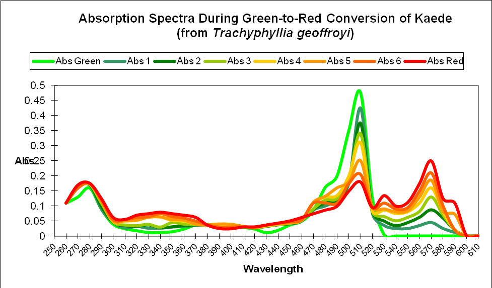
- A rearrangement of the chemical structure of a colorful protein by light. Depending upon the protein, photoconversion can increase or decrease fluorescence (in processes called photoactivation and photobleaching, respectively). Photoconversion can break proteins’ molecular bonds (as with Kaede and Eos fluorescent pigments) resulting in an irreversible color shift, or the molecule can be ‘twisted’ by light energy (a ‘hula twist’) where coloration reversal are possible depending upon the quality or quantity of light available. This process is known as photoswitching).
- Red Fluorescent Protein (RFP):
- Those pigments with an emission of ~570nm and above. Includes Ds-Red, Kaede and Chromo-Red pigments.
- Stokes Shift:
- The difference in the maximum wavelength of fluorescent pigment excitation light and the maximum wavelength of the fluoresced light (emission). For example, a pigment with an excitation wavelength of 508nm and an emission wavelength of 535nm would have a Stokes Shift of 27nm.
- Threshold or Coloration Threshold:
- The point at which pigment production is sufficient to make its fluorescence visually apparent. The term threshold generally refers pigment production, although, in some cases, it could apply to a light level where a pigment disappears (as in the cases of photobleaching, or photoconversion).
Types of Fluorescent Proteins
There are at least 9 described types of coral fluorescent proteins. Note: Pigment types, such as green or red might not be structurally similar to another green or red pigment in a different clade. These include:
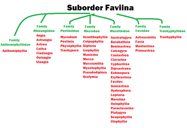
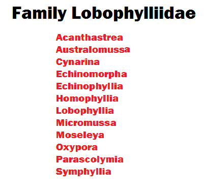
Figure 1. Coral taxonomy is in a state of constant flux, but this gives an idea of stony coral genera found in Suborder Faviina. Many of these animals contain Kaede or Clade D fluorescent proteins. Other coral genera contain them as well.
- Cyan Fluorescent Proteins (CFP) – Cyan pigments are blue-green pigments with a maximum emission of up to ~500 nm. The chromophore structure of a cyan pigment is very similar to that of a green fluorescent pigment.
- Green Fluorescent Proteins (GFP) – This group, by far, is the most numerous of the fluorescent proteins. The structure of green fluorescent chromophores is very similar to that of cyan fluorescent chromophores.
- Yellow Fluorescent Proteins (YFP) – An unusual type of fluorescent protein with maximum emission in the yellow portion of the spectrum. Rare in its biological distribution, YFP is found in a zoanthid and some specimens of the stony coral Agaricia. Personally, I’ve noted yellow fluorescence in a very few stony corals (Porites specimens) here in Hawaii while on night dives using specialized equipment to observe such colorations (see www.nightsea.com for details on this equipment).
- Orange Fluorescent Protein (OFP) – I’ve included this protein ‘type’ in an attempt to avoid confusion. OFP is used to describe a pigment found in stony coral Lobophyllia hemprichii and its name suggests a rather unique sort of protein. In fact, OFP is simply a variant of the Kaede-type fluorescent proteins.
- Red Fluorescent Proteins (RFP) – A group of proteins including several different subtypes (Kaede, Ds-Red and Chromo-Red). Typically, fluorescent emission is in the range of ~580 nm to slightly over 600 nm.
- Dronpa – A green fluorescent protein that loses fluorescent when exposed to blue-green light (~490 nm) but returns when irradiated with violet light at ~400 nm. Dronpa is a Clade D protein. Identified (so far) from a coral of Family Pectiniidae.
- Chromo-Red Proteins – A new classification (Alieva et al., 2008) of a single fluorescent pigment found in the stony coral Echinophyllia. This chromo-red pigment has some qualities of a non-fluorescent chromoprotein, but fluoresces at a maximum of 609 nm.
Corals Containing Clade D Fluorescent Proteins
As mentioned previously, almost 80 coral genera (at least 9 Families) contain Clade D (or Kaede-type) fluorescent proteins. Figures 1 – 4 (below) list these Genera by Family.
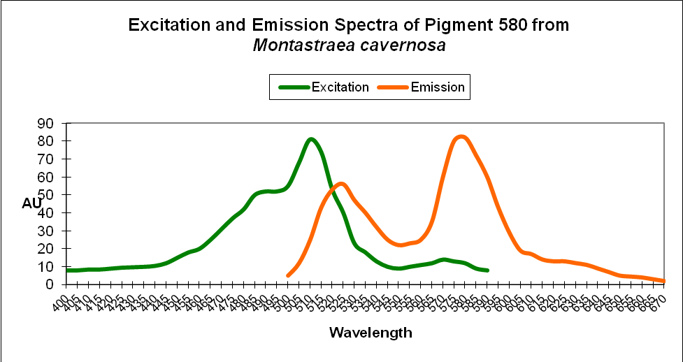
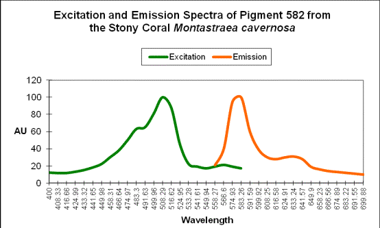
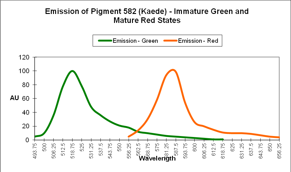
These fluorescent proteins are known by two names – the first is ‘Kaede-type proteins.’ Kaede is Japanese for ‘maple leaf’ refering to these proteins’ ability to transition from green to red color when irradiated with ultraviolet or strong blue light. These proteins are also known as ‘Clade D proteins’. Many Kaede-type (or Clade D) fluorescent proteins are easily discernible by their spectral emissions – there is a distinctive secondary shoulder at ~630nm when the fluorescence is in the orange/red portion of the spectrum. See Figure 5.
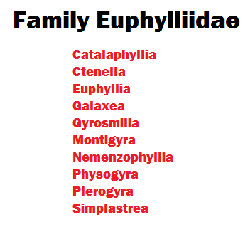
Figure 3. Family Euphylliidae contains many coral species popular among hobbyists. Some contain fluorescent proteins.
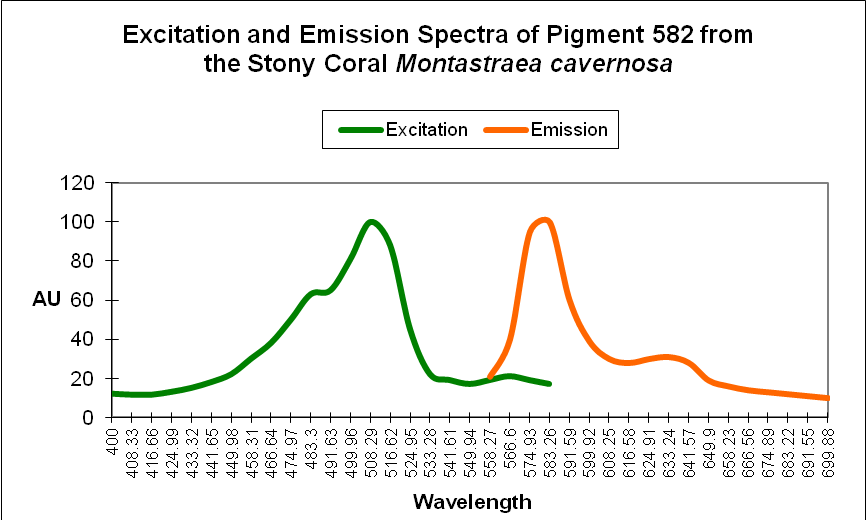
Figure 5. Clade D fluorescent proteins, when fully matured to orange/red, have a secondary emission (a ‘shoulder’) at ~630nm. ‘AU’ stands for arbitrary units.
Relationship of Clade D Proteins
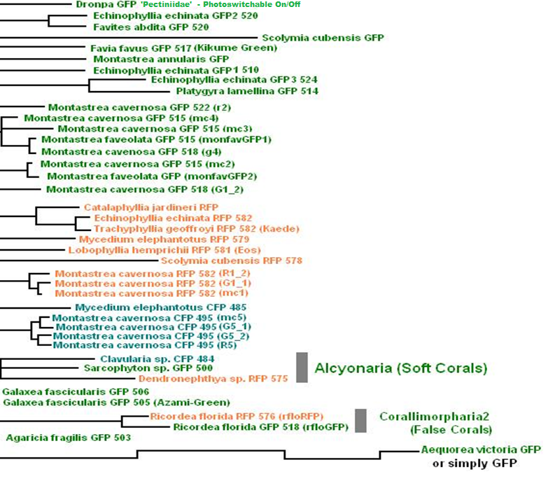
Figure 6. This phylogenetic tree lists Clade D fluorescent proteins, and their relationship to one another. The coral’s name is followed by ‘type’ of protein (CFP, GFP, and RFP) and maximum emission wavelength. In addition, some list the shorthand name used by researchers (such as rfloGFP found in Ricordea florida.
Figure 6 demonstrates the relationship of proteins found in various coral genera. In most cases, the coral species is listed, followed the type of protein (for example, GFP for ‘green fluorescent protein’, further refined in some cases by the maximum fluorescent emission wavelength (e.g., 520). The scientific shorthand is also listed (e.g., Kaede, Dronpa, mc5, etc.) The listing is also color-coded for easy recognition of maximum fluorescence color.
Clade D Corals
Now that we have the preliminaries out of the way, we can begin a detailed examination of information on Clade D fluorescent proteins. The following information is listed by coral genus in alphabetical order.
Agaricia
Common Name: Agaricia
Order: Scleractinia
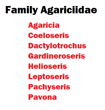
Family: Agariciidae
Agariciacorals are found only in the Atlantic Ocean, and are unlikely to be found in home aquaria. Proteins found in these corals fluorescein the blue-green, green, yellow-green, and yellow portions of the spectrum.
| Pigment | Emission | 2 | 3 | Excitation | 2 | 3 | Found in: |
|---|---|---|---|---|---|---|---|
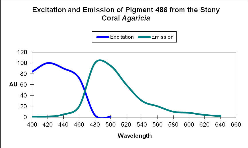
| P-486 | 486 | * | * | 426 | * | * | Agaricia sp. |
| P-497 | 497 | 527 | * | * | * | * | Agaricia sp. |
| P-503 | 503 | * | * | * | * | * | Agaricia fragilis |
| P-508-620 | 508-620 | * | * | * | * | * | Agaricia agaricites @ 60m |
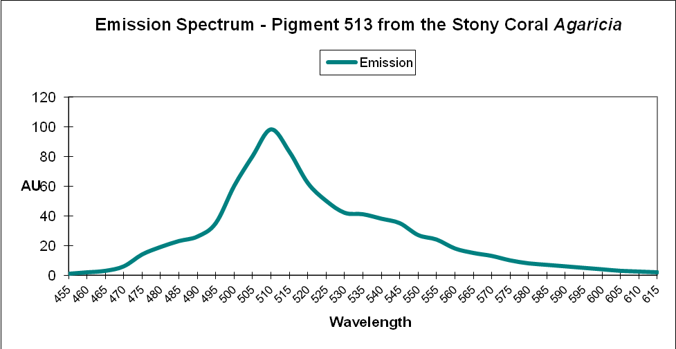
| P-513 | 513 | 545 | 490 | * | * | * | Agaricia sp. |
| P-515 | 515 | * | * | * | * | * | Agaricia sp. |
| P-542 | 542 | * | * | * | * | * | Agaricia undata @ 40m |
| P-557 | 557 | 600 | 545 | * | * | * | Agaricia sp. |
| P-565 | 565 | * | * | 490 | * | * | Agaricia humilis |
Catalaphyllia
Common Name: Elegance Coral, Elegant Coral
Order: Scleractinia
Family: Euphylliidae
Catalaphyllia specimens, commonly called Elegance or Wonder Corals, have been a mainstay in the hobby for years. The fleshy portion of the coral is usually fluorescent green, but tentacle tips can be orange. Some green fluorescent proteins in Catalaphyllia mature to red (the photoconvertible proteins described by researchers are likely found only in tentacles’ tips).
| Pigment | Emission | 2 | 3 | Excitation | 2 | 3 | Found in: |
|---|---|---|---|---|---|---|---|
| P-517 | 517 | * | * | 509 | * | * | Catalaphyllia jardinei |
| P-582 | 582 | * | * | 573 | * | * | Catalaphyllia jardinei |
Clavularia
Common Name: Clove Polyps
Order: Alcyonacea
Family: Clavulariidae
Only one fluorescent protein has been identified in Clavularia tissues, although there are obviously more. All soft corals’ pigments are closely related.
| Pigment | Emission | 2 | 3 | Excitation | 2 | 3 | Found in: |
|---|---|---|---|---|---|---|---|
| P-484 | 484 | * | * | 456 | * | * | Clavularia sp. |
Dendronephthya
Common Name: Tree coral, Strawberry coral
Order: Alcyonacea
Family: Nephtheidae
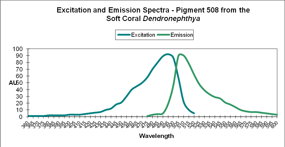
Figure 10. A green fluorescent protein found in the often non-photosynthetic soft coral Dendronephthya.
It may come as a surprise to many hobbyists, but some Dendronephthya specimens contain zooxanthellae. It is uncertain if Dendronephthya specimens containing fluorescent pigments possess zooxanthellae as well. Green and orange/red fluorescent proteins have been identified in these soft corals, and some have the ability to change from green to red upon exposure to ultraviolet, violet, or blue light.
| Pigment | Emission | 2 | 3 | Excitation | 2 | 3 | Found in: |
|---|---|---|---|---|---|---|---|
| P-508 | 508 | * | * | 494 | * | * | Dendronephthya |
| P-575 | 575 | * | * | * | * | * | Dendronephthya |
| P-575 | 575 | * | * | 557 | * | * | Dendronephthya |

Figure 11. The beautiful ‘Watermelon Chalice’ (Echinophyllia sp.). Photo courtesy Jason Fox Signature Corals (jasonfoxsignaturecorals.com).
Echinophyllia
Common Name: Chalice corals
Order: Scleractinia
Family: Lobophylliidae
Chalice corals are very popular, and for good reason. Their colors can be astounding and, not surprisingly, can be priced accordingly. Only a few fluorescent proteins are recognized in scientific literature, including 3 GFPs, one orange protein and a red one fluorescing at a maximum 0f 609nm. Interestingly, the P-582 protein found in Echinophyllia specimens is identical to another Kaede protein – the ‘original’ green-to-red transition protein found in Trachyphyllia geoffroyi. It is not unreasonable to assume that ultraviolet radiation and violet/blue light can cause the transition.
Coloration of the ‘Watermelon chalice’ can be explained by the green protein maturing to red. In areas of new growth, the coral would produce a GFP that would mature to red in older growth areas exposed to UV/violet/blue light. It is possible that the yellow ‘eyes’ in Figure 11 are due to an incomplete maturation where color mixing of green and red appear orange or yellow.
| Pigment | Emission | 2 | 3 | Excitation | 2 | 3 | Found in: |
|---|---|---|---|---|---|---|---|
| P-510 | 510 | * | * | * | * | * | Echinophyllia echinata |
| P-520 | 520 | * | * | * | * | * | Echinophyllia echinata |
| P-524 | 524 | * | * | * | * | * | Echinophyllia echinata |
| P-582 | 582 | * | * | * | * | * | Echinophyllia echinata |
| P-609 | 609 | * | * | * | * | * | Echinophyllia |
Favia
Common Name: Moon Coral
Order: Scleractinia
Family: Faviidae
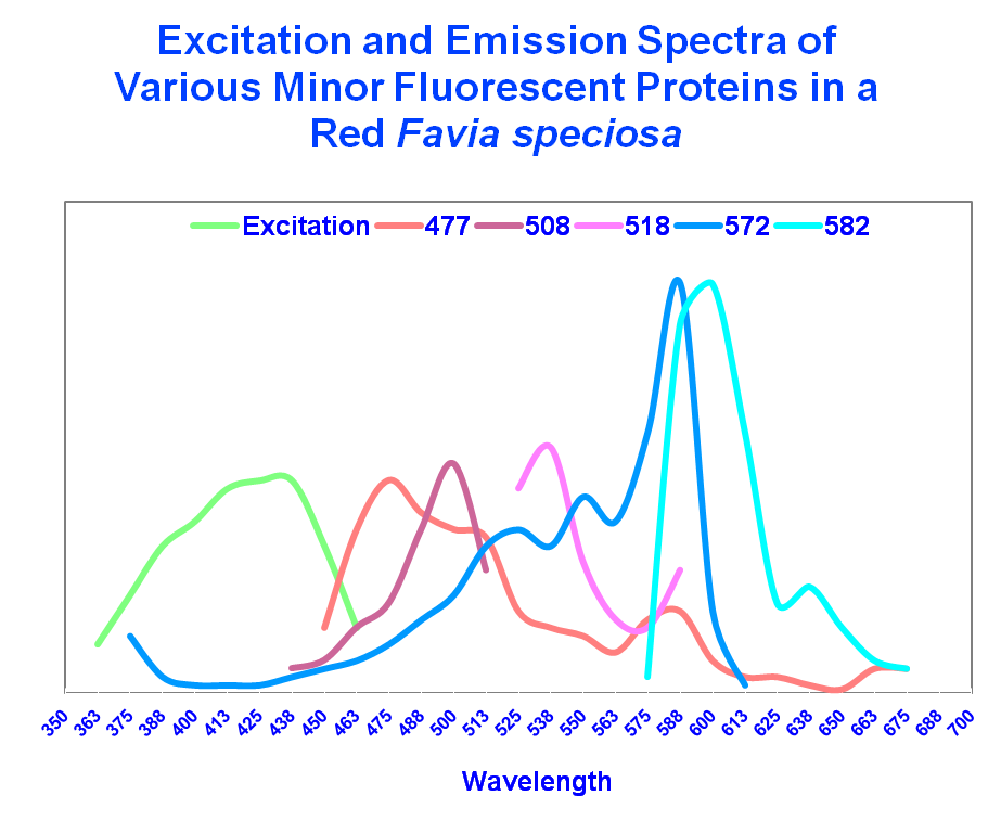
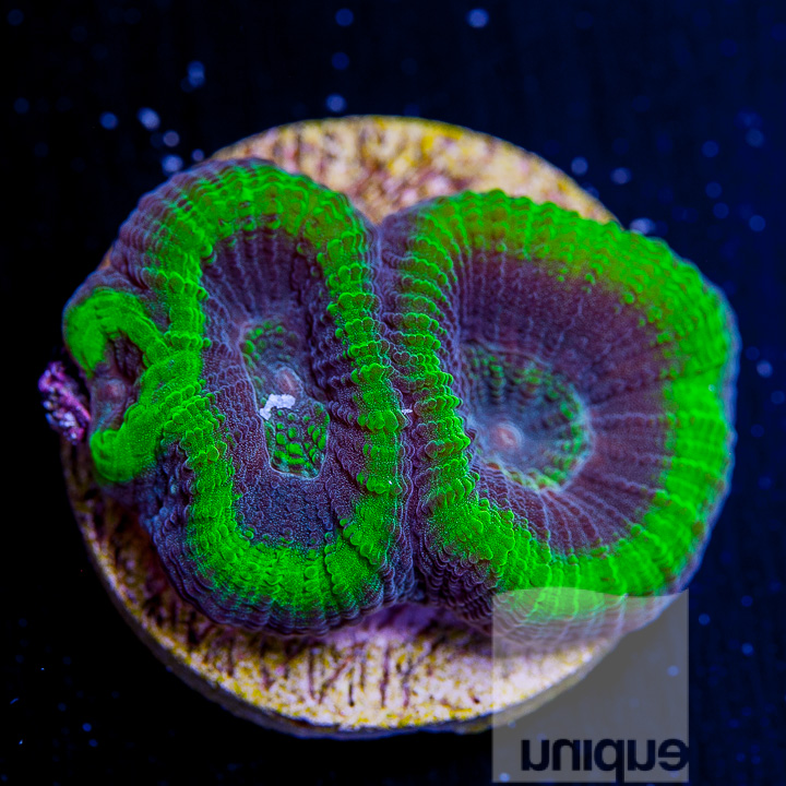
A Favia coral probably holds the record for possessing the most fluorescent colors (over 20, although they were not individually identified). Exposure to ultraviolet, violet, or blue light can cause some of the green proteins to mature to red ones. When color mixing is considered, the number of possible glowing color becomes staggering.
| Pigment | Emission | 2 | 3 | Excitation | 2 | 3 | Found in: |
|---|---|---|---|---|---|---|---|
| P-477 | 477 | * | * | 430 | * | * | Favia speciosa |
| P-507 | 507 | 536 | 575 | * | * | * | Favia fragum |
| P-508 | 508 | * | * | 430 | * | * | Favia speciosa |
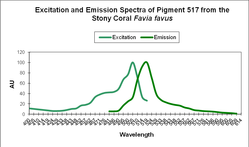
| P-517 | 517 | * | * | 507 | * | * | Favia favus |
| P-518 | 518 | * | * | 430 | * | * | Favia speciosa |
| P-572 | 572 | * | * | 430 | * | * | Favia speciosa |
| P-582 | 582 | * | * | 430 | * | * | Favia speciosa |
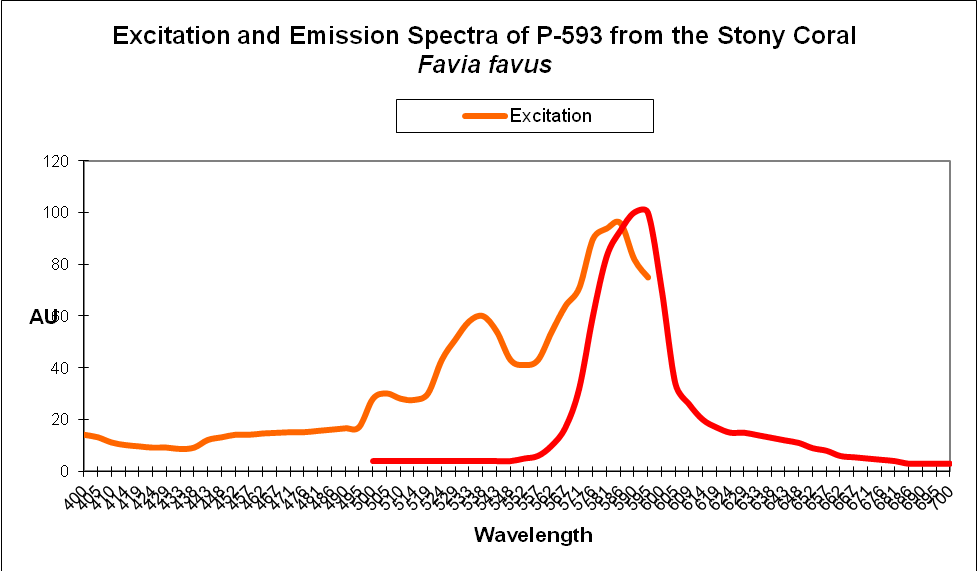
| P-593 | 593 | * | * | 583 | * | * | Favia favus |
Favites
Common Name: War coral, Honeycomb coral
Order: Scleractinia
Family: Merulinidae
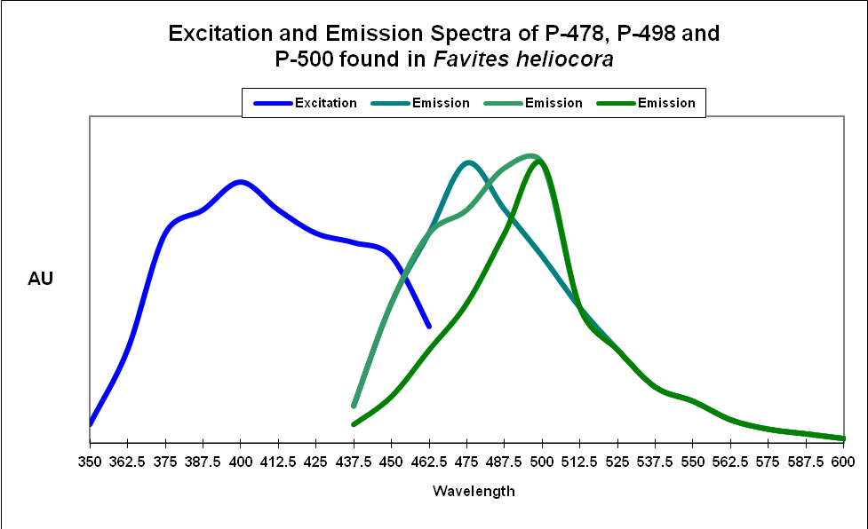
Figure 19. Excitation and emission wavelengths become blurred when fluorescence can be absorbed by another pigment. This Favites probably appears green.
Although science has recognized only blue-green and green fluorescence in Favites corals, aquarists know that orange/red fluorescent proteins are found in these corals as well.
| Pigment | Emission | 2 | 3 | Excitation | 2 | 3 | Found in: |
|---|---|---|---|---|---|---|---|
| P-478 | 478 | * | * | * | * | * | Favites heliocora |
| P-498 | 498 | * | * | * | * | * | Favites heliocora |
| P-500 | 500 | * | * | * | * | * | Favites heliocora |
| P-520 | 520 | * | * | * | * | * | Favites abdita |
Galaxea
Common Name: Galaxy coral
Order: Scleractinia Family: Euphyllidae
The highly aggressive coral Galaxea is known to contain several green fluorescent proteins.
| Pigment | Emission | 2 | 3 | Excitation | 2 | 3 | Found in: |
|---|---|---|---|---|---|---|---|
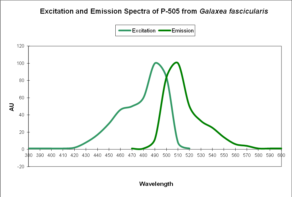
| P-505 | 505 | * | * | 492 | * | * | Galaxea fascicularis |
| P-505 | 505 | * | * | * | * | * | Galaxea fascicularis |
| P-506 | 506 | * | * | * | * | * | Galaxea fascicularis |
Lobophyllia
Common Name: Meat Coral
Order: Scleractinia
Family: Lobophyllidae
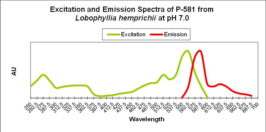
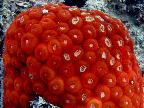
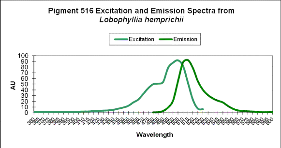
Some Lobophyllia hemprichii specimens contain the fluorescent protein known as ‘Eos’ (the goddess of dawn in Greek mythology). Eos is photo-activatible, and irreversibly transitions from green (emission at 516nm) to orange/red (581nm) when exposed to ultraviolet/violet light.
| Pigment | Emission | 2 | 3 | Excitation | 2 | 3 | Found in: |
|---|---|---|---|---|---|---|---|
| P-483 | 483 | * | * | 572 | 454 | 510 | Lobophyllia hemprichii (red) |
| P-515 | 515 | * | * | * | * | * | Lobophyllia hemprichii (red) |
| P-516 | 516 | * | * | * | * | * | Lobophyllia hemprichii |
| P-517 | 517 | Lobophyllia hemprichii | |||||
| P-519 | 519 | * | * | * | * | * | Lobophyllia hemprichii (red) |
| P-574 | 574 | 543 | Lobophyllia hemprichii (red) | ||||
| P-580 | 580 | * | * | * | * | * | Lobophyllia hemprichii (red) |
| P-581 | 581 | * | * | * | * | * | Lobophyllia hemprichii (red) |
Montastraea
Common Name: Boulder coral, Mountainous Star coral
Order: Scleractinia
Family: Montastraeidae
A recent revision to coral taxonomy has renamed Montastraea faveolata as Orbicella faveolata. Whatever the coral genus, the fluorescent proteins found in Montastraea species appear to be closely related.
Although corals of the genus Montastraea are found in the Atlantic and Pacific, only Atlantic species have been examined for fluorescent proteins. It is not unreasonable, in my opinion, to expect Pacific Montastraea species to contain similar, if not identical, fluorescent proteins.
| Pigment | Emission | 2 | 3 | Excitation | 2 | 3 | Found in: |
|---|---|---|---|---|---|---|---|
| P-480 | 480 | * | * | * | * | * | Montastraea annularis |
| P-484 | 484 | 499 | * | * | * | * | Montastraea annularis |
| P-486 | 486 | * | * | * | * | * | Montastraea annularis |
| P-503 | 503 | 479 | 578 | * | * | * | Montastraea annularis |
| P-510 | 510 | 479 | * | * | * | * | Montastraea annularis |
| P-515 | 515 | * | * | 505 | * | * | Montastraea annularis |
| P-517 | 517 | 551 | 483 | * | * | * | Montastraea annularis |
| P-518 | 518 | 575 | * | * | * | * | Montastraea annularis |
| P-486 | 486 | * | * | 440 | * | * | Montastraea cavernosa |
| P-505 | 505 | * | * | * | * | * | Montastraea cavernosa |
| P-510 | 510 | * | * | 440 | * | * | Montastraea cavernosa |
| P-510-520 | 510-520 | * | * | 440 | * | * | Montastraea cavernosa |
| P-514 | 514 | * | * | * | * | * | Montastraea cavernosa |
| P-517 | 517 | * | * | 504 | * | * | Montastraea cavernosa |
| P-519 | 519 | * | * | ~505 | * | * | Montastraea cavernosa |
| P-575 | 575 | ~630 | * | ~525 | ~570 | * | Montastraea cavernosa |
| P-516 | 516 | * | * | 506 | ~480 | * | Montastraea cavernosa (=mc2/3/4 – see P-515) |
| P-582 | 582 | * | * | * | * | * | Montastraea cavernosa (G1_1) |
| P-518 | 518 | * | * | * | * | * | Montastraea cavernosa (G1_2) |
| P-518 | 518 | * | * | * | * | * | Montastraea cavernosa (g4) |
| P-495 | 495 | * | * | * | * | * | Montastraea cavernosa (G5_1) |
| P-495 | 495 | * | * | * | * | * | Montastraea cavernosa (G5_2) |
| P-582 | 582 | 630 | * | 508 | 572 | * | Montastraea cavernosa (mc1) |
| P-515 | 515±3 | * | * | 505±3 | * | * | Montastraea cavernosa (mc2/3/4) |
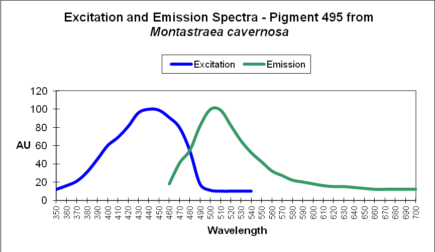
| P-495 | 495 | * | * | 435 | * | * | Montastraea cavernosa (mc5) |
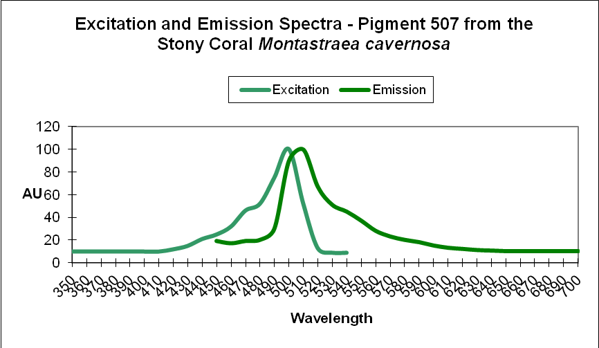
| P-507 | 507 | * | * | 495 | * | * | Montastraea cavernosa (mc6) |
| P-580 | 580 | 520 | * | 508 | 572 | * | Montastraea cavernosa (mcavRFP) |
| P-582 | 582 | * | * | * | * | * | Montastraea cavernosa (R1_2) |
| P-522 | 522 | * | * | * | * | * | Montastraea cavernosa (r2) |
| P-495 | 495 | * | * | * | * | * | Montastraea cavernosa (R5) |
| P-510-623 | 510-623 | * | * | * | * | * | Montastraea cavernosa @ 40m |
| P-534 | 534 | 593 | * | * | * | * | Montastraea cavernosa @ 60m |
| P-486 | 486 | * | * | 440 | * | * | Montastraea faveolata |
| P-510-520 | 510-520 | * | * | 440 | * | * | Montastraea faveolata |
| P-515 | 515 | * | * | * | * | * | Montastraea faveolata |
| P-582 | 582 | * | * | 571 | * | * | Montastrea cavernosa |
Mycedium
Common Name: Peacock coral, Plate coral
Order: Scleractinia
Family: Pectiniidae
At least one coral in Family Pectiniidae contains the fluorescent protein known as Dronpa (a disappearing Ninja according to folklore). This green fluorescent protein photo-bleaches when exposed to blue-green light (~490nm). Upon exposure to violet light (~400nm), the green fluorescence returns. Since many lamps generate both wavelengths, it is uncertain how broad spectrum light affects coloration in aquarium corals.
| Pigment | Emission | 2 | 3 | Excitation | 2 | 3 | Found in: |
|---|---|---|---|---|---|---|---|
| P-485 | 485 | * | * | * | * | * | Mycedium elephantotus |
| P-579 | 579 | * | * | * | * | * | Mycedium elephantotus |
Platygyra
Common Name: Brain coral
Order: Scleractinia
Family: Faviidae
| Pigment | Emission | 2 | 3 | Excitation | 2 | 3 | Found in: |
|---|---|---|---|---|---|---|---|
| P-514 | 514 | * | * | * | * | * | Platygyra lamellina |
Plesiastrea
Common Name: Pineapple brain coral
Order: Scleractinia
Family: Scleractinia incertae sedis (rough translation: ‘scleractinia family uncertain’)
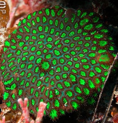
I find it ironic that a system based on order is in a state of constant chaos, where coral families are re-sorted according to the latest information. In any case, if Plesiastrea’s Scleractinian Family is in doubt, it seems certain that the fluorescent protein found in these corals falls into Clade D.
Quite a few fluorescent proteins have been identified in Plesiastrea corals including CFP’s, GFP’s, yellow-green proteins, in addition to orange and super-reds.
| Pigment | Emission | 2 | 3 | Excitation | 2 | 3 | Found in: |
|---|---|---|---|---|---|---|---|
| P-472 | 472 | * | * | * | * | * | Plesiastrea verispora (blue morph) |
| P-473 | 473 | * | * | * | * | * | Plesiastrea verispora (blue morph) |
| P-473 | 473 | * | * | * | * | * | Plesiastrea verispora (green morph) |
| P-477 | 477 | * | * | * | * | * | Plesiastrea verispora (blue) |
| P-479 | 479 | * | * | * | * | * | Plesiastrea verispora (blue morph) |
| P-479 | 479 | * | * | * | * | * | Plesiastrea verispora (green morph) |
| P-482 | 482 | * | * | * | * | * | Plesiastrea verispora (blue) |
| P-485 | 485 | * | * | * | * | * | Plesiastrea verispora (blue morph) |
| P-485 | 485 | * | * | * | * | * | Plesiastrea verispora (green morph) |
| P-488 | 488 | * | * | * | * | * | Plesiastrea verispora (blue morph) |
| P-489 | 489 | * | * | * | * | * | Plesiastrea verispora (blue morph) |
| P-489 | 489 | * | * | * | * | * | Plesiastrea verispora (green morph) |
| P-492 | 492 | * | * | * | * | * | Plesiastrea verispora (blue) |
| P-495 | 495 | * | * | * | * | * | Plesiastrea verispora (blue morph) |
| P-495 | 495 | * | * | * | * | * | Plesiastrea verispora (green morph) |
| P-497 | 497 | * | * | * | * | * | Plesiastrea verispora (blue morph) |
| P-497 | 497 | * | * | * | * | * | Plesiastrea verispora (green morph) |
| P-501 | 501 | * | * | * | * | * | Plesiastrea verispora (blue morph) |
| P-503 | 503 | * | * | * | * | * | Plesiastrea verispora (green morph) |
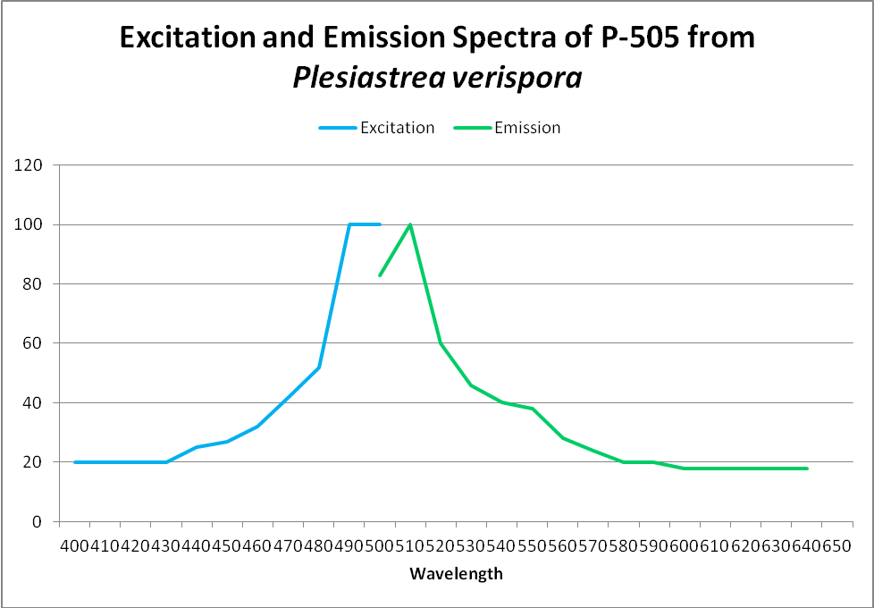
| P-505 | 505 | * | * | 492 | * | * | Plesiastrea verispora |
| P-508 | 508 | * | * | * | * | * | Plesiastrea verispora (green morph) |
| P-511 | 511 | * | * | * | * | * | Plesiastrea verispora (green morph) |
| P-512 | 512 | * | * | * | * | * | Plesiastrea verispora (blue morph) |
| P-512 | 512 | * | * | * | * | * | Plesiastrea verispora (green morph) |
| P-515 | 515 | * | * | * | * | * | Plesiastrea verispora (green morph) |
| P-515 | 515 | * | * | * | * | * | Plesiastrea verispora (green morph) |
| P-518 | 518 | * | * | * | * | * | Plesiastrea verispora (green morph) |
| P-540 | 540 | * | * | * | * | * | Plesiastrea verispora (blue morph) |
| P-540 | 540 | * | * | * | * | * | Plesiastrea verispora (green morph) |
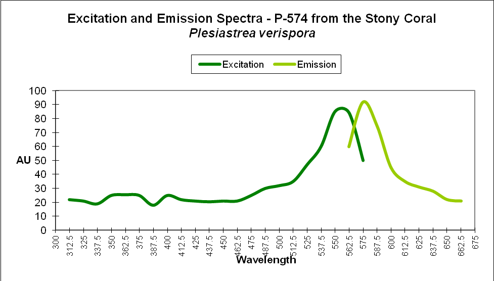
| P-574 | 574 | 550 | * | * | * | * | Plesiastrea verispora |
| P-580 | 580 | * | * | * | * | * | Plesiastrea verispora (blue morph) |
| P-580 | 580 | * | * | * | * | * | Plesiastrea verispora (green morph) |
| P-620 | 620 | * | * | * | * | * | Plesiastrea verispora (blue morph) |
| P-620 | 620 | * | * | * | * | * | Plesiastrea verispora (green morph) |
Ricordea
Common Name: False coral, Hairy Mushroom coral
Order: Corallimorpharia
Family: Ricordeidae
Ricordeaspecies have a number of devotees and for good reasons – their ease of maintenance and beautiful colorations make them excellent candidates for reef aquaria. Scientists have shown aquarists that fluorescent proteins within these corals can transition from green to orange/red. It is supposed that blue light causes this transformation.
| Pigment | Emission | 2 | 3 | Excitation | 2 | 3 | Found in: |
|---|---|---|---|---|---|---|---|
| P-510 | 510 | * | * | * | * | * | Ricordea florida |
| P-513 | 513 | * | * | * | * | * | Ricordea florida |
| P-515 | 515 | * | * | * | * | * | Ricordea sp. |
| P-517 | 517 | 574 | * | 506 | 566 | * | Ricordea florida |
| P-517 | 517 | * | * | 506 | * | * | Ricordea florida |
| P-518 | 518 | * | * | 508 | 475 | * | Ricordea florida |
| P-518 | 518 | * | * | * | * | * | Ricordea florida |
| P-518 | 518 | * | * | * | * | * | Ricordea florida |
| P-520 | 520 | * | * | * | * | * | Ricordea florida |
| P-573 | 573 | 510 | * | * | * | * | Ricordea florida |
| P-574 | 574 | 517 | * | 506 | 566 | * | Ricordea florida |
| P-576 | 576 | * | * | * | * | * | Ricordea florida |
| P-587-590 | 587-590 | * | * | * | * | * | Ricordea florida (mouth) |
Sarcophyton
Common Name: Leather coral, Toadstool coral
Order: Alcyonacea
Family: Alcyoniidae
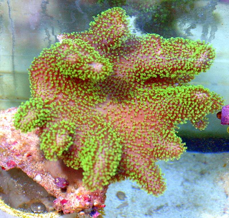
Sarcophytoncorals have been a mainstay of the hobby for many years. They are easy to maintain in captivity and, in good water flow, offer a dynamic sense to an aquarium otherwise full of rigid, stony corals. In addition, their green fluorescence adds beauty to any reef aquarium.
| Pigment | Emission | 2 | 3 | Excitation | 2 | 3 | Found in: |
|---|---|---|---|---|---|---|---|
| P-500 | 500 | * | * | * | * | * | Sarcophyton sp. |
Scolymia
Common Name: Button coral, Doughnut coral
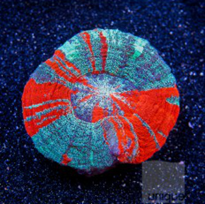
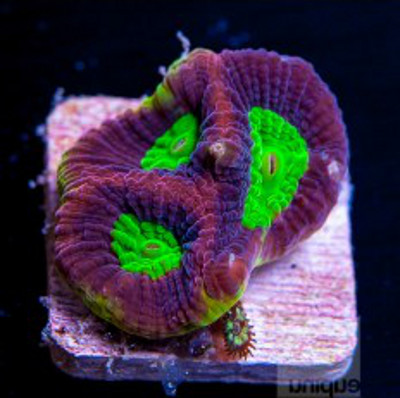
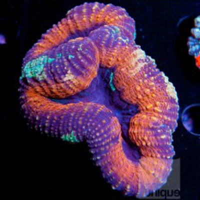
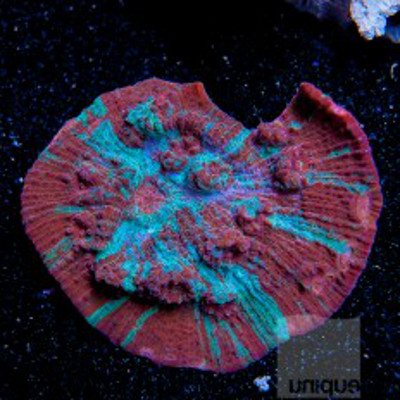
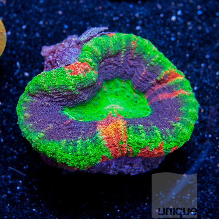
Figure 38. This Scolymia appears to contain at least 3 fluorescent proteins. Courtesy uniquecorals.com.
Order: Scleractinia
Family: Mussidae
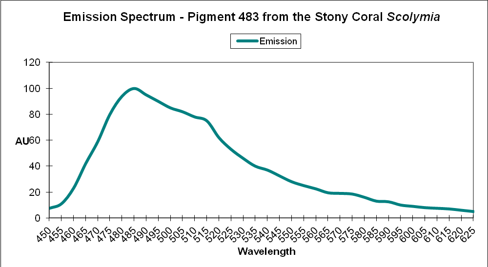
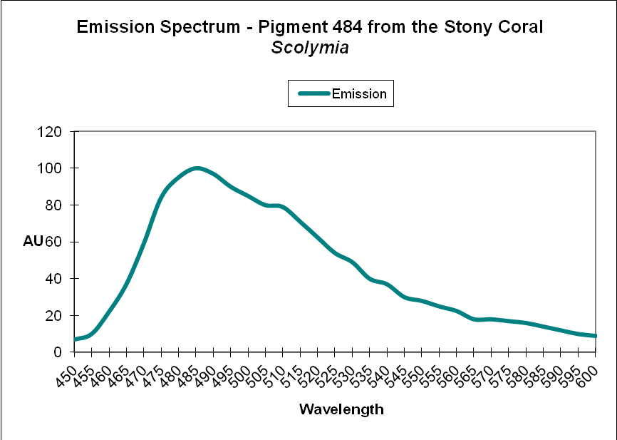
Colorful Scolymia specimens are popular – and often expensive. Their fluorescent proteins range in coloration from blue-green to orange-red. Some consider specimens in this genus to be a bit touchy in captivity if they are not fed regularly.
| Pigment | Emission | 2 | 3 | Excitation | 2 | 3 | Found in: |
|---|---|---|---|---|---|---|---|
| P-483 | 483 | 511 | * | * | * | * | Scolymia sp. |
| P-483 | 483 | 511 | 576 | * | * | * | Scolymia sp. |
| P-484 | 484 | 512 | * | * | * | * | Scolymia sp. |
| P-484 | 484 | 512 | 576 | * | * | * | Scolymia sp. |
| P-506 | 506 | * | * | 497 | * | * | Scolymia cubensis 1 |
| P-506 | 506 | * | * | 497 | * | * | Scolymia cubensis 2 |
| P-515 | 515 | * | * | * | * | * | Scolymia sp. |
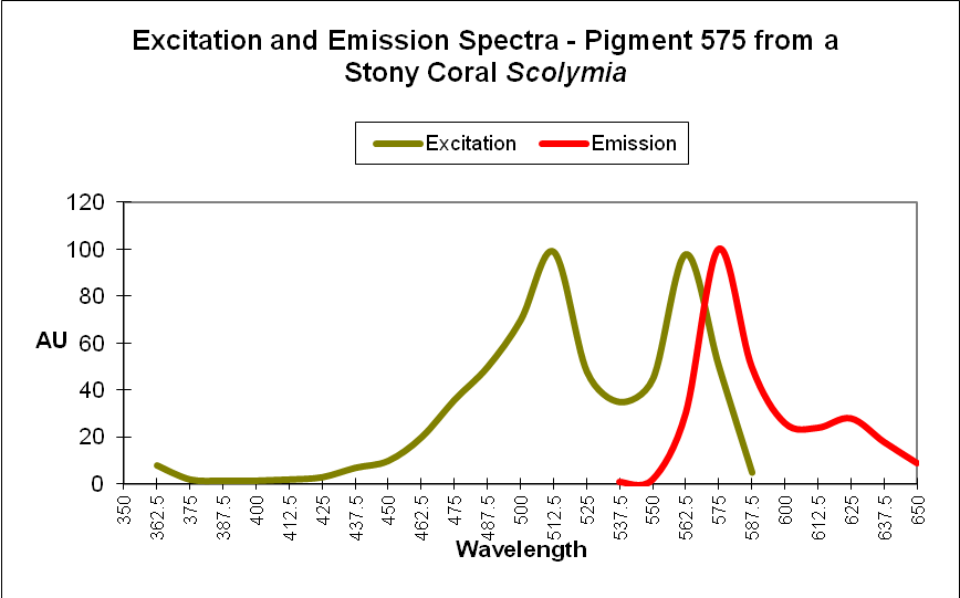
| P-575 | 575 | 630 | * | 520 | * | * | Scolymia sp. |
| P-578 | 578 | * | * | * | * | * | Scolymia cubensis |
Trachyphyllia
Common Name: Open Brain Coral
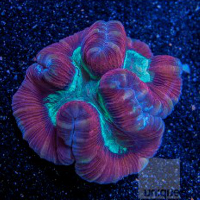
Figure 43. The ‘original’ Kaede protein found in Trachyphyllia geoffroyi. Photo Courtesy of uniquecorals.com.
Order: Scleractinia
Family: Trachyphylliidae
The Family Trachyphylliidae is composed of one genus – Trachyphyllia. It was from this coral that the Kaede group of fluorescent proteins was originally described. ‘Kaede’ is Japanese for ‘maple leaf’, referring to its green to red transition when exposed to ultraviolet, violet, or blue light. Note that the intermediate transition from green to red can produce yellow or orange intermediates.
| Pigment | Emission | 2 | 3 | Excitation | 2 | 3 | Found in: |
|---|---|---|---|---|---|---|---|
| P-518 | 518 | * | * | * | * | * | Trachyphyllia geoffroyi |
| P-582 | 582 | * | * | 558 | * | * | Trachyphyllia geoffroyi |
Fluorescent Proteins Likely to Belong to Clade D, but Unconfirmed
Based strictly on genera, these corals probably contain Clade D fluorescent proteins and may act similarly to others in the group.
Genus: Colpophyllia
Family: Mussidae
| Emission | 2 | 3 | Excitation | 2 | 3 | Found in: |
|---|---|---|---|---|---|---|
| 515 | * | * | * | * | * | Colpophyllia natans |
Genus: Diploria
Family: Mussidae
| Emission | 2 | 3 | Excitation | 2 | 3 | Found in: |
|---|---|---|---|---|---|---|
| P-486 | 486 | * | ~448 | * | * | Diploria labyrinthiformis |
| P-517 | 517 | 575 | * | * | * | Diploria strigosa |
Genus: Hydnophora
Family: Merulinidae
| Emission | 2 | 3 | Excitation | 2 | 3 | Found in: |
|---|---|---|---|---|---|---|
| P-492 | 492 | * | * | 443 | * | Hydnophora grandis |
Genus: Manicina
Family: Mussidae
| Emission | 2 | 3 | Excitation | 2 | 3 | Found in: |
|---|---|---|---|---|---|---|
| P-487 | 487 | 515 | 475 | * | * | Manicina areolata |
Genus: Mycetophyllia
Family: Mussidae
| Emission | 2 | 3 | Excitation | 2 | 3 | Found in: |
|---|---|---|---|---|---|---|
| P-515 | 515 | * | * | * | * | Mycetophyllia lamarckiana |
| P-515 | 515 | * | * | ~494 | * | Mycetophyllia sp. |
Genus: Leptoseris
Family: Agariciidae
Mikhail Matz (one of the world’s leading experts on coral fluorescence) argues that this protein was isolated from this deep-water specimen through not use of water but a polar solvent instead. This fact suggests the protein may be different from all others.
| Emission | 2 | 3 | Excitation | 2 | 3 | Found in: |
|---|---|---|---|---|---|---|
| P446(?) | 446 | * | * | 380 | * | Leptoseris fragilis |
Genus: Pavona
Family: Agariciidae
The only fluorescent protein identified so far from Pavona fluoresces in the green portion of the spectrum. However, some Pavona specimens are fluorescent orange. Is this an example of photoconversion from green to orange?
Effects of pH
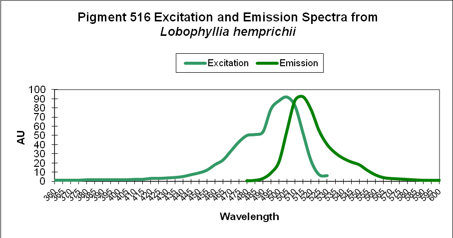
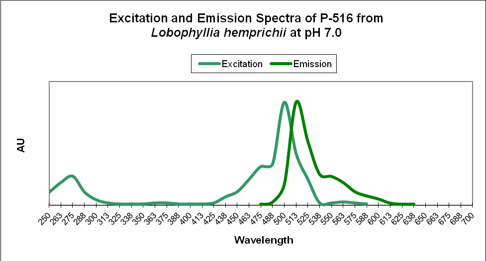
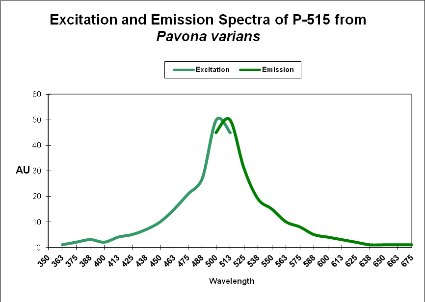
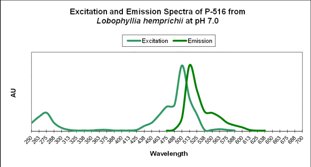
pH is known to affect the absorption and fluorescent characteristics of some proteins. These changes tend to be slight, but it is not unreasonable to believe (supported by anecdotal evidence) that large swings in apparent color could be caused by extreme pH modulations (caused by an overdose of carbon dioxide or kalkwasser). See Figures 48 and 49.
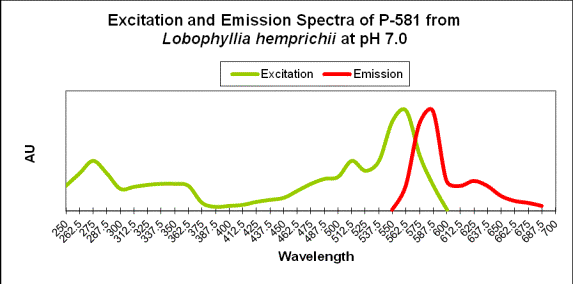
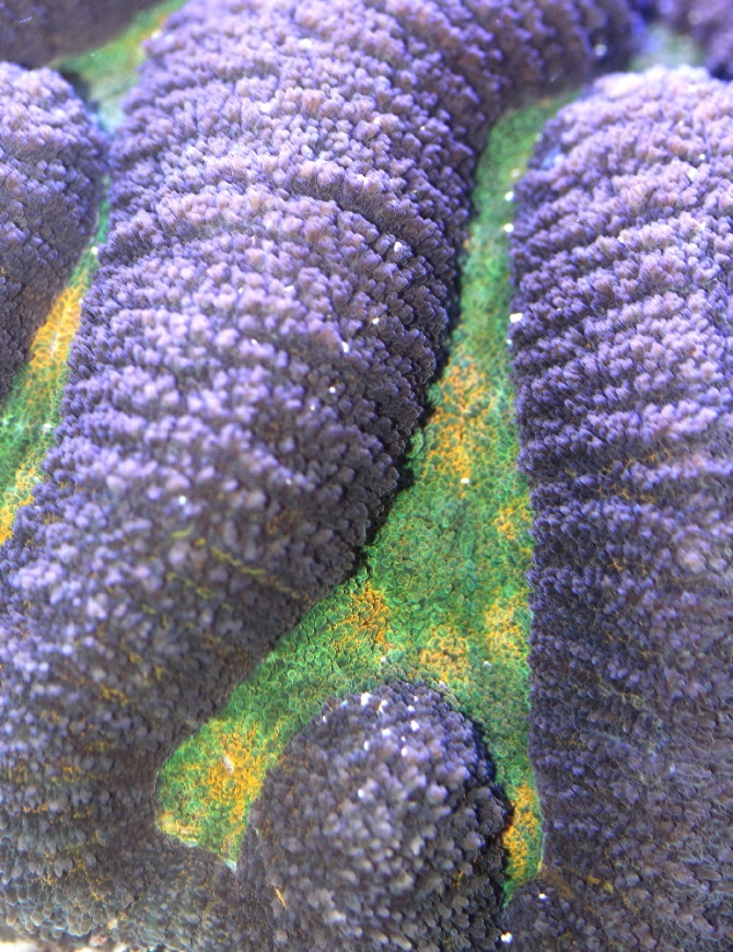
Figure 50. Note that yellow-green light excites the red fluorescence in this Lobophyllia coral. Note also the low pH – this affects the absorption and emission of the protein.
Discussion
Proteins of Clade D are found in over 80 coral genera (at least 9 families). Evidence suggests many are photo-convertible by ultraviolet radiation and violet/blue light (although blue LEDs – producing practically no ultraviolet radiation and little violet light) are known to induce expression and maintain coloration in many corals.)
Table 24. Conversion of green fluorescent proteins to the Kaede type orange/red pigments has been observed when the animal is exposed to Ultraviolet-A radiation (UV-A), and violet/blue wavelengths. Care should be taken when experimenting with dosages of UV-A in attempts to promote coloration. Most plastic ‘splash guards’ are transparent to at least some UV-A wavelengths and it is usually not necessary to deliberately increase UV-A levels in order to induce production of pigments. With this said, there is some anecdotal information (based on my personal observations) that color of at least some pigments might increase in at least some coral species when UV radiation is slightly increased.
| From | To | Host | Clade/Pigment | Activator |
|---|---|---|---|---|
| P-505 | 508/572 | Montastraea cavernosa | D | ? – But most likely blue light |
| P-505 | 506/566 | Ricordea florida | D | ? – But most likely blue light |
| P-508 | 575 | Dendronephthya sp. | D | Blue Light @ 488nm |
| P-508 | 575 | Dendronephthya sp. | D | UV-A at 366nm |
| ~516 | 582 | Montastraea cavernosa | D | Depth- light -related? |
| P-516 | 581 | Lobophyllia hemprichii | D | UV @ 390nm/Violet Light ~400nm |
| P-517 | 574 | Ricordea florida | D | UV/Violet Light |
| P-517 | 580 | Montastraea annularis | D | UV/Violet Light |
| P-517 | 593 | Favia favus | D | UV & Violet (350-420nm) |
| P-518 | 582 | Trachyphyllia geoffroyi | D | UV – Violet Light (350-410nm) |
| P-519 | 580 | Montastraea cavernosa | D | UV – Violet Light |
However, many blue-green and green fluorescent proteins are present in conditions of low light, suggesting their presence is not related to light intensity (or perhaps generated in conditions of very low light).
To confound matters, at least one colorful protein (Dronpa) is known to photo-bleach when exposed to blue-green light at 490nm, but the fluorescence returns if exposed to blue light. Fortunately (for aquarists), researchers have found that Dronpa is not closely related to many other Clade D proteins (see Figure 6).
pH
Schreiner et al. (2011) report the red fluorescence of the Eos protein (from Lobophyllia sp.) decreases when exposed to an acidic pH (<7.0), while the green fluorescent emission is unaffected (this in contrast to others’ observations). Apparently, some GFPs are affected by pH while others are not. When looking at the fluorescent protein phylogenetic tree, we see the red Eos protein is clustered with several other red fluorescent proteins, including those from Trachyphyllia, Catalaphyllia, Mycedium, Echinophyllia, and Scolymia. We might expect the red fluorescence of these pigments to be affected similarly by low pH.
An acidic pH would be an unusual condition within a reef aquarium, but it is possible, perhaps as a result of an overdose of carbon dioxide through use of a calcium reactor or similar device. On the other hand, a high pH (caused by an overdose of kalkwasser) might affect apparent coloration as well.
To summarize, light (particularly violet and/or blue) seems to be the environmental trigger for inducing the production of fluorescent proteins (ultraviolet light can, in many cases, also cause their production. However, the use of LEDs and their almost total lack of UV production strongly suggest UV is not necessary.) Another factor – pH – is known to slightly alter the fluorescent emission of these ‘pigments’.
Many Clade D proteins’ fluorescent emissions change as the protein ages (assuming the exposure to violet/blue light continues. This is seen dramatically in the ‘watermelon chalice’ as the green margin edges matures and turns red). The transition from green to red is responsible for an almost unlimited number of intermediate colors. However, recall that many Clade D proteins do not make a transition from green to red.
Many of the Clade D proteins cloned for use in biomedical research are stable at higher (human body temperatures) suggesting they are relatively unaffected by temperature modulations.
Our understanding of coral fluorescent proteins is due largely to experiments conducted in the biomedical fields and we have come a long way since corals’ colors were attributed to photopigments such as peridinin (erroneously thought at one time to lend a brick-red color to corals). However, early observations that corals tended up to ‘color up’ when exposed to blue light have been proven to be essentially correct. With the recent revelation that is possible to provide at least as much (or in some circumstances, more) blue light than corals could expect to ‘see’ in nature, further experiments can proceed in examining the effects of altered spectra on coral photosynthesis and expression of coloration (see here for details: http://www.advancedaquarist.com/2013/11/aafeature ).
Acknowledgements
Taxa are classified according to World Register of Marine Species (WoRMS) www.marinespecies.org and are correct as of early October 2013. Any errors are mine.
Many thanks to Joe Caparatta at Unique Corals (www.uniquecorals.com) for supplying many of the photographs.
References
- Scheiner, L., M. Huber-Lang, M. Weiss, H. Hohmann, M. Schmolz, and E. Schneider, 2011. Phagocytosis and digestion of pH-sensitive dye (Eos-FP) transfected E. coli in whole blood assays from patients with severe sepsis and septic shock. J. Cell. Commun. Signal, 5(2):135-144.



0 Comments