Most corals and all giant clams (tridacnids) contain symbiotic algae, which they rely upon for much of their nutritional needs. When these algae, commonly known as zooxanthellae, receive adequate illumination they can produce far more food than is required for their own survival, growth, and reproduction, and this excess can be given to the host animal they reside in. However, under a range of adverse conditions these animals may lose some or almost all of their complement of zooxanthellae and become pale in color, or even completely clear letting their white skeleton show through in the case of stony corals. When this loss of zooxanthellae and the various pigments associated with them occurs and a host loses its color, the condition is appropriately called bleaching. It can affect small areas of a coral or tridacnid at times, but in severe cases it can affect the whole host.
Here’s a good video on YouTube:
http://www.youtube.com/watch?v=60jof35WuAo
(and many others) if you’d like to see examples and the effects of large-scale bleaching on reefs.

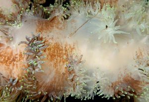
Elegance coral in the process of bleaching. You can see strands of zooxanthellae and mucus being expelled from the corals mouths.
Both corals and tridacnids oftentimes recover from bleaching in the wild, but those housed in aquariums oftentimes don’t unless action is taken to remedy the situation. Therefore, it’s important to understand what can cause this condition, the different types of bleaching, and what to do if it occurs.
Causes of Bleaching
There are several things that can cause bleaching to occur at some scale or another, which I’ll cover below. However, over-illumination (light shock) and/or unacceptably high temperatures are the primary culprits in aquariums, and in the wild, too. So, I’ll start with these.
Corals and tridacnids can protect themselves from over-illumination in a number of ways, and can to a large degree adapt to any increases in light intensity over time. But, if the amount of light (especially UV light) that a coral or tridacnid receives is simply too high, or increases faster than the organism can adapt to the change, the result can be biochemical changes that lead to an unhealthy overload of the photosynthetic processes carried out by the zooxanthellae and cellular damage (see Borneman 2001 for more on this). Unacceptably high temperatures can also adversely affect photosynthetic processes, so something has to change when light intensity goes up too much and/or too quickly and/or temperatures get too high.
If any of these occur a reduction in the number of zooxanthellae that corresponds to the severity of the situation is the typical result. It may be only a small decrease in numbers in some situations, but in most cases the size of the population may drop dramatically. In fact, almost every last algal cell may disappear, leaving their host just about empty. That’s only in extreme cases though, as there’s usually at least a small population of zooxanthellae that’s held onto, which can later begin to reproduce and repopulate a host’s tissues if conditions improve. In such situations a host may capture zooxanthellae from the environment to rebuild its complement at a later time, too.
Under normal conditions in reef environments the length of days, the average daily cloud cover, and water temperatures typically change very slowly through the seasons and corals and tridacnids can adapt to these regular changes. However, there are times when temperatures increase more and/or faster than usual, and there are times when decreased wind/wave activity allows more light to penetrate deeper into waters, too.
Wave activity can break up incoming sunlight to some degree, and can also keep sediments stirred up at least a bit in shallow waters, which also block some incoming sunlight. However, no wind means no waves, and no waves means more light making it through the surface while suspended sediments simultaneously settle to the bottom allowing even more light fall upon whatever is below. This can lead to light shock and then to bleaching as more than the normal dose of light reaches corals and tridacnids. Likewise, bleaching can also occur in an aquarium if either is subjected to illumination that is more intense than what they’re used to without being given adequate time to adapt and/or if the temperature ever gets too high. Do note that if given plenty of time for adjustment, it is practically impossible to give a tridacnid too much light, though. All species can live in very shallow waters and tolerate full sunlight without problems, and I’m pretty sure your aquarium lights are nowhere near as bright as the sun.
Regardless, this is why you absolutely must give corals and tridacnids time to adapt to your lighting system unless you are positive that your lights are dimmer than what the specimen is already adapted to. The same goes for temperature, as you should never run an aquarium at an unacceptably high temperature, or allow temperatures to spike if your air conditioner should ever fail.
So, light shock and high temperatures are the main culprits, but corals and tridacnids can also bleach as a result of under-illumination, or being kept in water that isn’t warm enough. They can also bleach due to a lack of required nutrients, unacceptable salinities, poisoning by metals, too much red light, the use of some medications, or being over-stressed for some reason (ex. Duquesne & Coll 1995, Brown 1997, Braley 1998, and Borneman 2001). And, as if that’s not a long enough list, disease-related bleaching has been reported to occur in some corals (Kushmaro et al. 1996 and Rosenberg & Loya 1999) and was also suggested by Norton et al. (1995) as a probable candidate for some cases of tridacnid bleaching. However, while the bacteria Vibrio shiloi was positively identified as the pathogen in some cases of coral bleaching, no specific bacteria or other microorganism has been identified as the cause of tridacnid bleaching. Still, there’s strong evidence that it’s possible, so something specific may be found in the future.
Knop (1996) also suggested that some cases of bleaching may result from an inability of a host or its zooxanthellae to produce certain substances when kept in an aquarium. Zooxanthellae do produce a number of required pigments, and they’re also the likely source of the substances that a host uses in order to produce other pigments and UV-screening compounds themselves (Borneman 2001). So, if for some reason things go wrong for either or both partners, these may be difficult or impossible to make.
Types of Bleaching
Bleaching can generally be seen in three different forms. There’s generalized bleaching and localized bleaching that affect corals and tridacnids, then there’s central bleaching that affects only tridacnids. So, let’s look at each of these and why they’re thought to occur.
 Generalized bleaching is seen as a relatively uniform loss of color over an entire coral/coral colony or the entire mantle of a tridacnid (the soft, extendable tissue that protrudes from the shell). It can range in severity from a slight lightening to a complete loss of color, and it’s the most likely type to lead to a specimen’s death. This is most likely to occur as a result of light shock, elevated temperatures, too little light and/or nutrients (starvation), too high or low salinity, or some sort of poisoning. Essentially anything that affects the whole host.
On the other hand, localized bleaching is quite different and can be seen as patches of lightening that may occur anywhere on a host. This can range from a slight lightening to a complete loss of color, and it’s unlikely that unacceptable lighting or temperatures, or poisoning, etc. are the cause. Instead, localized bleaching may be the result of an infection of some sort, as mentioned above, or some sort of physical injury/damage to a restricted area of a host’s tissues. It may also occur if a portion of the specimen is heavily shadowed by an overhang, a coral branch or branches, etc.
If such bleaching occurs as a reaction to being shadowed, it should remain restricted roughly to the area that isn’t receiving sufficient light. But, if it’s due to an infection (or something else), it may start as a small area and then spread over larger portions of the specimen at times. Still, in some cases it shows up in one spot for no apparent reason and doesn’t spread much if any at all, as I’ve seen occur many times on tridacnid mantles.
As long as it doesn’t spread over much of the host, localized bleaching doesn’t seem to have any noticeable effect on the overall fitness of tridacnids. I’ve seen numerous specimens with small areas of bleaching that still grew normally, and didn’t seem to be bothered at all. Oddly enough, it tends to be permanent too, at least for tridacnids. In the case of corals, it also may persist, or spread, or go away over time.
Of course, some localized bleaching may be the first signs of generalized bleaching, too. While generalized bleaching typically affects most or the whole host simultaneously, at times it can start as a single or several patches that increase in size over time. So, you need to keep a close watch on a specimen and watch for progression even if only a small area bleaches.
Then there’s central bleaching, which is seen as a loss of color in the central area of a tridacnid’s the upper mantle, particularly on any relatively flat portions of it. It can also range in severity from a slight lightening to a complete loss of color, and is most likely the result of over-illumination. I say this because only part of a specimen is typically affected and this area is roughly perpendicular to incoming light, being exposed even if the shell is only partially open and the mantle is retracted. However, Knop (1996) also suggests that it may be due to a reduced ability to produce sunscreens, as mentioned above. Regardless, central bleaching is far less likely to cause the death of a tridacnid. But, if action isn’t taken to correct to situation that brought it on, it may spread over the rest of the mantle.

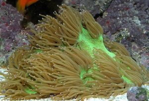
An example of a coral suffering from localized bleaching, probably related to the injured areas at the edge of the skeleton.
This can be a tough one to be sure about though, as many tridacnids have areas of their mantles that are devoid of color and look rather translucent which are not the result of bleaching. In particular, specimens of Tridacna gigas often have a substantial amount of color-free mantle tissue, and T. derasa sometimes has colorless, round spots dotted over its mantle, too. Really, all of the species can have some colorless areas at times, particularly in the central area of the mantle, so don’t hastily jump to conclusions if you happen to see any light spots. Such areas may not be signs of trouble.
The same goes for branch tips on many stony corals. It’s common for the very ends of the branches of an Acropora specimen, or some other small-polyp coral to be white in color, but again, in this case it’s normal. Still, if you see an obvious overall lightening or increase in the size and/or abundance of colorless areas over a relatively short period of time then you do need to take action.

An example of a tridacnid suffering from localized bleaching for unknown reasons. I was told this white patch was permanent and always stayed the same size.

Note that it is common for T. gigas to have significant translucent areas on its mantle, which are not a form of bleaching.

It is also common for T. derasa to have translucent, roughly circular patches all over its mantle, which are not a form of bleaching.
What to Do
Due to the fact that there are so many things that can lead to bleaching, it can be quite difficult to identify its specific cause at times. However, by starting with the basics, and using your head, there’s a good chance you can find and fix the problem.

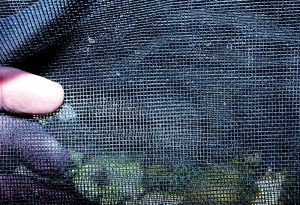
Screening material can come in handy if a situation arises when light intensity needs to be reduced over a specimen.
To start, it is imperative that your water quality is up to par, and you should immediately correct anything that is outside acceptable ranges. Aside from that, the most obvious is to check the temperature. While reef aquariums are best kept in the range of 77° to 82°F, many hobbyists may run their tanks a little higher or lower than this. However, if your air conditioner ever quits or a heater gets stuck on, etc. temperatures can easily rise a few degrees and that’s all it takes. Thus, if something in your tank bleaches due to unacceptable temperatures, you need to do whatever it takes to get it into that 77° to 82º range as quickly as possible. Even if your tank normally runs higher than 82º, I think you’ll be better off reducing the temperature to 82º or less until everything has recovered (if it does).
The next thing is deciding if a coral or tridacnid is getting too much light. If you’ve gotten a new specimen of some sort and it starts to bleach after you’ve acclimated it to your aquarium, and you feel confident that it’s not due to any of the things above, then you may not have given it adequate time to fully adapt to your lighting. You typically won’t know what lighting conditions the specimen was living under before it arrived at a store, so there’s always the possibility that your lighting may actually be significantly brighter than what it’s used to.
The easiest thing to do in such a situation is to use one or more sheets of screening material to reduce the light the specimen is receiving. This stuff is available at most any hardware/home improvement stores, and you can cut out pieces of material that shade the specimen in trouble by placing it on a glass top, or using clothes pins to affix a strip over the top of one area of the tank, without shading everything else in the tank that’s doing fine. The best thing to do is to cut down the light using a few overlaid pieces of screen and then remove one piece at a time over a period of a couple of weeks. This will also allow you to leave the specimen wherever you placed it, rather than move it around over and over, which can be particularly bad for tridacnids.
Likewise, a specimen may be light shocked when you change the bulbs in your lighting system, especially if you wait too long to do so. Bulbs get dimmer as time goes by, but if you replace them often enough there shouldn’t be too much difference between the output of the old and the new. However, I imagine lots of hobbyists keep bulbs running longer than they should, which means that when they do get replaced, there will be a greater difference between the output of the old bulbs and their replacements. Possibly enough difference in output to lead to bleaching. So, if a specimen starts to bleach shortly after a bulb replacement, then you can guess that the change was very likely the cause, and should take appropriate action such as the use of shade cloth.
It’s not very likely, but waiting way too long between water changes, or adding large amounts of carbon to a system can lead to light shock, too. Over time aquarium water can become increasingly yellow in color due to the accumulation of substances collectively called gelbstoff or gilvin, which are produced primarily by the decay of organic materials. These can absorb significant amounts of light (Bingman 1996), and if their concentration is rapidly reduced the amount of light hitting a specimen can rapidly increase.
Water changes can reduce the concentration of yellowing substances, and so can the use of activated carbon, and/or using a skimmer. Thus, you shouldn’t do large, infrequent water changes that would instantaneously and significantly reduce their concentration and potentially let enough additional light hit a specimen to cause it to bleach. Likewise, you shouldn’t use large amounts of activated carbon and replace it infrequently as this could potentially lead to bleaching, too. Skimmers, on the other hand, tend to be run continuously, so it’s unlikely that simply running a skimmer will ever cause a dramatic change in the concentration of yellow substances. However, if you decide to add a skimmer where there wasn’t one before, or upgrade to a bigger/better skimmer, keep in mind that this may also quickly reduce yellowing compounds.
If you think that a specimen has started to bleach due to such a reduction in yellowing substances, try the shade cloth. Bleaching under these circumstances is still light shock, albeit due to other reasons, so you’ll need to reduce the light and hope that things get better.
Bleaching due to under-illumination can occur as a result of either never having enough lights over your aquarium to keep a particular specimen healthy long-term, or it could happen if your lights were okay when they were new, but have gone too long without being replaced. As I said, they get dimmer over time, and if you wait long enough they just might get too dim.
In such cases the solution is obvious. You can either upgrade your lighting system so that it’s adequate, or get new bulbs to replace old ones. Be careful though, because if your lights are so dim that a specimen is starving/bleaching and you suddenly increase the intensity dramatically, you may end up making things worse rather than better.
Frankly, I can’t imagine that bleaching will ever occur in an aquarium due to a lack of nutrients unless there are no fishes in the tank. But, if you’re convinced that it’s happening because of this, you obviously need to add some. There are several quality products available that contain various sorts of plankton or plankton-substitutes, which can be used to help ailing specimens, and to feed perfectly healthy tanks, too.
Then there’s the possibility of disease. Unfortunately, if bleaching occurs due to an infection of some sort, there isn’t much that you can do about it. I say this because there wouldn’t be any way for a typical hobbyist to identify what type of microorganism was responsible for the bleaching. So, you’d potentially have to treat a specimen in a quarantine tank (I never add drugs to a display tank) with a variety of antibiotics, hoping that one of them kills off the infection. To make things worse, a treatment could be successful, but you might not know it until several weeks later. The offensive microbes could be dead, but the color of the specimen would still take a while to return. Again, this would be a very difficult thing to treat considering the unknowns and efforts involved. Fortunately, this is also very unlikely to occur.
Lastly, we get to stress-induced bleaching that may have nothing to do with any of these other things. Getting bagged up and shipped across the world, or just from one side of the country to another can be very stressful for specimens, and they may respond by bleaching. An annoying fish can stress out a specimen, parasitic snails can stress tridacnids, a physical injury of some sort, or even something as simple as an unacceptable current may have the same effect in some cases, even if everything else is fine. So, aside from everything covered above, keep an eye on a specimen to see if it is being bothered by obnoxious tankmakes, or is being blasted by strong currents, etc., and remedy the situation immediately if you find anything wrong.
In any case, regardless of what you decide is the probable cause, keep in mind that a host is highly stressed when bleached (with the possible exception of small-scale localized or central bleaching), and will very likely benefit from some added food. Planktonic/particulate foods may increase a specimen’s chances of recovering, as they can help to make up for the lack of food produced by the zooxanthellae. So, even if you usually don’t feed your tank, I’d recommend that you do so at least until everything seems to be healthy and looking good.
References and sources for more information
- Asada, K. and M. Takahashi. 1987. Production and scavenging of active oxygen in photosynthesis. In: Kyle, D.J., C.B. Osmond, and C.J. Arntzen (eds.) Photoinhibition. Elsevier, Amsterdam. 307pp.
- Borneman, E. 2001. Aquarium Corals – Selection, Husbandry, and Natural History. Microcosm/T.F.H. Publications, neptune City, NJ. 464pp.
- Braley, R.D. 1998. Report to GBRMPA on results of research done under marine parks permit no.G92/137. Aquasearch: http://www.aquasearch.net.au/aqua/long.htm
- Brown, B.E. 1997. Coral bleaching: causes and consequences. Coral Reefs 16:S129-S138.
- Duquesne, S.J. and J.C. Coll. 1995. Metal accumulation in the clam Tridacna crocea under natural and experimental conditions. Aquatic Toxicology 32:239-253.
- Fatherree, J.W. 2006. Giant Clams in the Sea and the Aquarium. Liquid Medium, Tampa. 227pp.
- Knop, D. 1996. Giant Clams: A Comprehensive Guide to the Identification and Care of Tridacnid Clams. Dahne Verlag, Ettlingen, Germany. 255pp.
- Kushmaro, A., Y. Loya, M. Fine, and E. Rosenberg. 1996. Bacterial infection and coral bleaching. Nature 380:396.
- Leggat, W., B.H. Buck, A. Grice, and D. Yellowlees. 2003. The impact of bleaching on the metabolic contribution of dinoflagellate symbionts to their giant clam host. Plant, Cell and Environment 26:1951-1961.
- Norton, J.H., H.C. Prior, B. Baillie, and D. Yellowlees. 1995. Atrophy of the zooxanthellal tubular system in bleached giant clams Tridacna gigas. Journal of Invertebrate Pathology 66:307-310.
- Osmond, C.B. 1981. Photorespiration and photoinhibition; some implications for the energetics of photosynthesis. Biochemica Biophysica Acta 639:77-98.
- Rosenberg, E. and Y. Loya. 1999. Vibrio shiloi is the etiological (causative) agent of Oculina patagonica bleaching: General implications. Reef Encounters 25:8-10


0 Comments