

Strains of the pathogenic dinoflagellate Pfiesteria piscicida of the family Pfiesteriaceae have been implicated in mass kills of fin- and shell-fish predominantly in estuarine environments, but their permissive range extends to full-strength seawater (35 parts per thousand; ppt; o/oo; ~1.026 SG) and 33oC (91oF; Fig 10.; Burkholder et al. 1995; Springer et al. 2002). However, most nearshore marine fish are euryhaline (Wu & Woo 1983) which likely hunt in estuaries. Pfiesteriaceae’s aptitude for causing horrific disease in any finfish epitomises the broadest host range, while their toxins have poisoned humans (Glasgow et al. 1995; Grattan et al. 2001). The family comprises over 20 species, where laboratory-cultured Luciella masanensis ingested a variety of plankton and human and fish erythrocytes (Jeong et al. 2007), whereas Pfiesteria shumwayae has instigated epizootics in both brackish and freshwater recirculating systems (Moestrup et al. 2014).
Similar life stages are adopted by Stoeckeria algicida of the family Thoracosphaeraceae, whose tri-flagellated planozygote bioremediates the toxigenic red tide raphidophyte Heterosigma akashiwo (Jeong et al. 2005; Gottschling et al. 2012; Lun et al. 2017). Strains of this species cannot feed directly from fish, yet they can ingest their red blood cells (Jeong et al. 2007a; Jeong et al. 2007) while manmade eutrophy contributes to outbreaks (Burkholder et al. 1995). Thoracosphaeraceae thus benefit from coinfections with Pfiesteriaceae.
Taxonomic Context
Members of the families Pfiesteriaceae and Thoracosphaeraceae are classified within the kingdom Chromista, the subkingdom Harosa, the infrakingdom Alveolata, the phylum Myzozoa, the subphylum Dinozoa, the infraphylum Dinoflagellata, the class Dinophyceae, and the order Thoracosphaerales (WoRMS 2022a; WoRMS 2022b).
The presence of fish and their waterborne excreta triggers the transmutation of inactive amoebae or the germination of cysts to form the infective dinospores of P. piscicida and P. shumwayae (Burkholder & Glasgow 1995; Litaker et al. 2002). These life phases develop toxigenic competencies, where binary fission culminates in cyst germination while sexual reproduction follows gamete production (Litaker et al. 2002). Toxins fortuitously anaesthetise before they induce necrotic ulcerations which creates waterborne suspensions of cells, their debris, and enriched phosphate (Noga et al. 1996) which fortify biochemical stimuli. Stasis or erythrocytic, somatic cellular, and phytoplanktonic ingestion safeguards the environmental persistence of these dinoflagellates (Burkholder & Glasgow 1995; Burkholder et al. 1995). Spores of Pfiesteriaceae attach to fish using tow filaments whilst their pedunculate baskets extract the contents of cells (Fig 2.; Schnepf & Elbrachter 1992; Jeong et al. 2007).
Clinical Signs
Anticipate perceptible surface zoning and aggregation in contaminated recirculating systems conceivably due to their current or archaic photosynthetic competency and phytoplanktivory (Gwang Hoon et al. 2013). Dinoflagellate feeding, toxin-mediated necrosis, and secondary infections participate in lesion development (Schnepf & Elbrachter 1992; Burkholder et al. 1995; Noga 2000; Jeong et al. 2007). Teleosts become narcotised and descend to the bottom whilst being literally eaten alive, whereas nearby gelatinous matrices are redolent of sexual reproduction via gametic fusion (Burkholder & Glasgow 1997; Litaker et al. 2002).
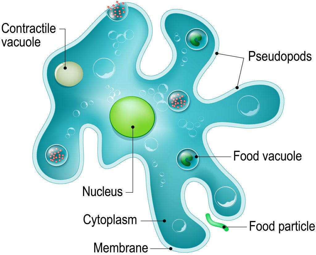
Fig 1. A representation of a unicellular protozoa of the genus Amoeba which belong to an extraordinarily diverse group of some 50 genera that inhabit moist/aquatic environments. Their symbiotic bacteria cause sporadic diseases in humans, while some that live in saltwater inhabit shells (Fig 12.; Schulz et al. 2015).
Diagnosis
Brightfield microscopy combined with clinical pathology assists in putative confirmation, where the images in Burkholder and Glasgow (1997a; 1997) and Litaker and collaborators (2002) prove invaluable.
Life Cycle
Flagellated motile Pfiesteria piscicida remain haploid for most of their life with only one copy of each chromosome (1n), and like strains of the aetiological agents of marine velvet (Amyloodinium ocellatum), their infectious free-swimming stage is referred to as a dino- or zoo-spore (Fig 6.).
Numerous microbes are exploited for sustenance in the absence of fish (Mallin et al. 1995; Litaker et al. 2002) although they instantaneously swarm onto a host when teleosts arrive (Schnepf & Elbrachter 1992; Burkholder & Glasgow 1997; Litaker et al. 2002; Jeong et al. 2007). Here we present the outcomes of inconsistent findings insofar as unlike Burkholder and Glasgow’s research published in 1997, Litaker and colleagues’ study from 2002 failed to identify amoebae and cysts with scales akin to those of diatoms of the phylum Chrysophyta.
Asexual Reproduction
Well-fed dinospores swim in slow circles, round up, discard their flagella, and fall to the bottom as vegetative cysts, whereas conjoined zoospores emerge, separate, and become free-swimming after germinative rupture. Malnourished dinospores transform into resting cysts that settle and remain viable for several months, whereas stressed P. piscicida temporarily encyst then undergo internal division or germinate to form solitary spores (Litaker et al. 2002).
One form of Sexual Reproduction occurs via the fusion of gelatinous matrix-bound haploid (1n) visually indistinguishable isogamous and virtually isogamous gametes (Burkholder & Glasgow 1997; Litaker et al. 2002). Surprisingly, nutritional dearth does not induce gametogenesis. Conjugative pairs of gametic spores spiral in the water where the leading retains feeding competency while the other is towed. The anterior of the trailing fuses with the posterior of the former whereupon plasmogamy spawns a sizeable heart shaped, binucleate, di-flagellated planozygote within 24 hours which becomes ellipsoidal when karyogamy fuses its nuclei. Discharge of its flagella precedes its transformation into a nonmotile spherical hypnozygote whose mitosis results in a tetraploid nucleus (4n) whereafter meiosis I and II generate two diploid (2n) meiocytes from which four haploid sister gametes are formed (Fig 3.). Mature hypnozygotes germinate into four daughter dinospores within 48 hours (Litaker et al. 2002).
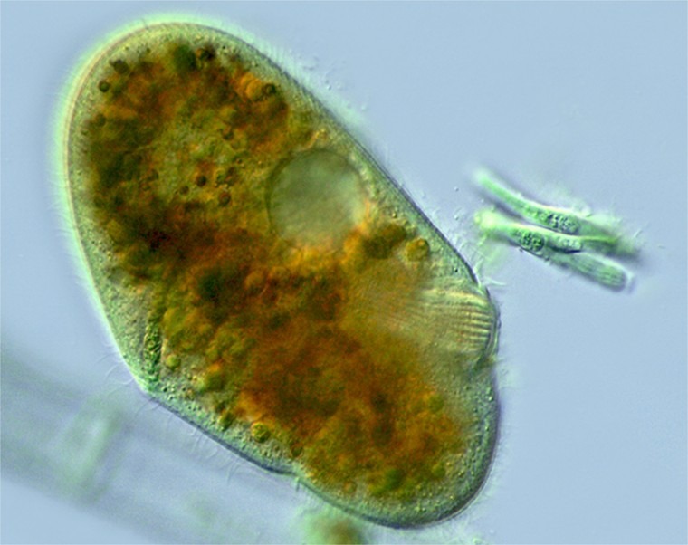
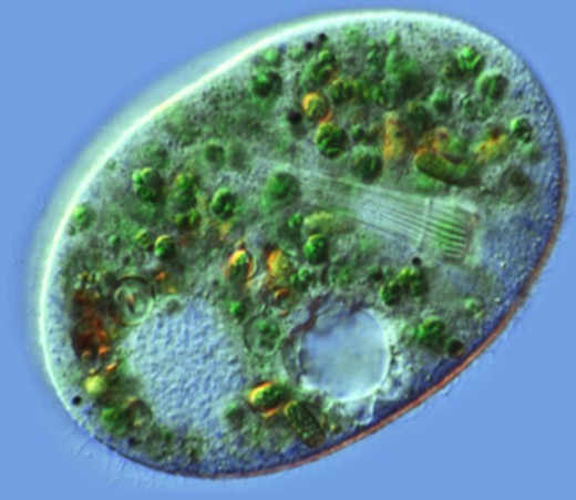
Fig 2. Micrographs of chromists of the genus Nassula (Ciliophora: Nassophorea) featuring their basket-like cytopharyngeal tubes (CYTs) lined with rod-like structures called nematodesmata (Hausmann & Hülsmann 2010) whose extremities resemble the pedunculate baskets of Pfiesteria. Images courtesy of Antonio Guillén©, Proyecto Agua, Flikr.com
Non-pathogenic or -toxic strains of P. piscicida rarely transform into amoebae (Burkholder et al. 2001a; Burkholder et al. 2001; Burkholder et al. 2005) while these conclusions were deduced from studying a benign species (Samet et al. 2001; Burkholder & Glasgow 2002). A later study observed the development of amoebae and chrysophyte-like cysts whose observations were consistent with Steidinger and colleagues from 1996, Burkholder and Glasgow from 1997, and Marshall and allies published in 2000 (Burkholder & Glasgow 1997a; Burkholder & Glasgow 2002). The experimenters witnessed isogamous and perceptibly distinct anisogamous gametes (Burkholder & Glasgow 2002) where toxic flagellates predominated throughout fish survival while amoebae consumed their carcasses (Burkholder et al. 1995; Burkholder & Glasgow 1997a).
A Complex Life Cycle
Fish-derived chemical cues trigger cyst germination into toxic motile amoebae or nontoxic dinospores (Burkholder & Glasgow 1997a), whereas anomalous dips in temperature of around 15oC may elicit outbreaks of sizeable lobose toxigenic amoebae. Lobosa is now a subphylum which was a class replaced by Tubulinea (WoRMS 2022c). Nontoxic dinospores swarm onto and attach via tow filaments to piscine hosts and commence to phagocytose their cells including erythrocytes where they transform into planozygotes or toxic zoospores (Burkholder & Glasgow 1997a). Such attacks are exacerbated by enriched dissolved inorganic phosphorus (DIP; phosphate; Burkholder et al. 1995) common to fish-only recirculating or partial flow-through systems, which in turn fuels the growth of their phytoplanktonic prey and stimulates the production of isogamous gametes (Burkholder & Glasgow 1997a). All flagellated life stages feed using vacuuming stalked baskets including gametes (Hausmann & Hülsmann 2010) which look like the extremities of the phagocytic cytopharyngeal tubes of other chromists (Fig 2.).
Breaches in the skin of narcotized fish fortify chemical signals as they are consumed almost entirely by toxic dinospores, while their ensuing mortality triggers the formation of vestige-devouring lobose or cyst-forming star-like rhizopodial amoebae. Zooplanktonic amoebae are sustained on other plankton or lie dormant in sediment, and toxic dinospores produce anisogamous gametes which evolve into planozygotes, whereas elevated DIP maintains gametes as persistent plankton (Steidinger et al. 1996; Burkholder & Glasgow 1997a).
Several yet more complex life stages emerge which were presented in Burkholder and Glasgow in 1997(a), yet their interrelations are too intricate to be included here, therefore figure 5. illustrates a simplistic and incomplete overview.
Harmful algal bloom (HAB) mitigating strategies are discussed below, while studies isolated significantly diverse microbial consortia comprising Pfiesteria-like organisms and over 30 other species dominated by a-Proteobacteria. Nonetheless, such communities were not unlike those of the holobionts of most HABs (Fig 4.; Alavi et al. 2001).
Contrasting Evidence
A series of epizootics caused by Pfiesteria and Pfiesteria-like microbes were reported in the summer and autumn of 1996 and 1997 in the Pocomoke River in Maryland and North Carolina’s tributaries of Chesapeake Bay. The most conspicuous of which was a disease of Atlantic menhaden (Brevoortia tyrannus; Fig 11.; Burkholder & Glasgow 1997; Blazer et al. 2016) whose ulcerations were colonized by fungi. Previous research had emphasized the opportunistic nature of mycoses; however, hyphae did not extend beyond lesion boundaries (Fig 8.; Noga et al. 1996, cited in Burkholder & Glasgow 1997; Noga et al. 1996, cited in Blazer et al. 2016).
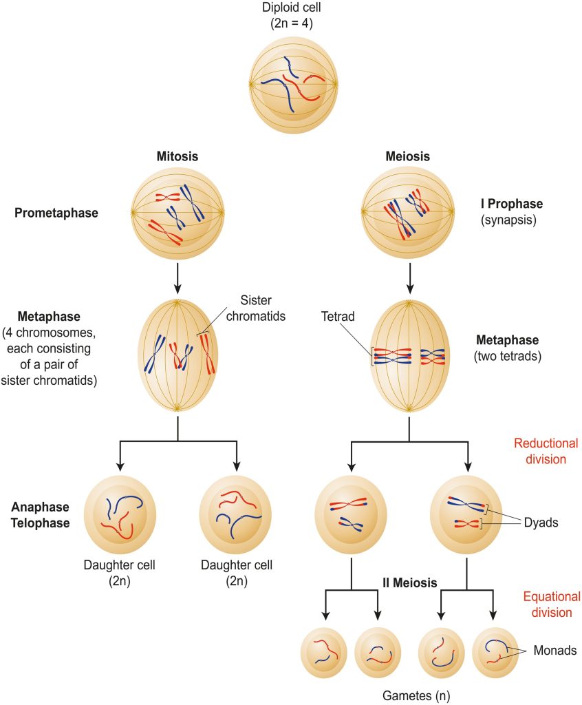
Fig 3. Diploid cellular binary fission: mitosis versus meiosis where meiosis II generates haploid gametes.
The skin lesions of stressed and/or immunocompromised fish may be instigated by opportunistic pathogens from several phyla, and menhaden collected in the summer of 1997 and 1998 from Chesapeake Bay, exhibited haemorrhagic fungal wounds or deep ulcerations. Histological analyses revealed chronic inflammatory responses surrounding deep penetrating hyphae that pervaded skeletal muscles and pierced viscera (Fig 9.; Blazer et al. 1999; Blazer et al. 2016).
Outbreaks of Asian epizootic ulcerative syndrome (EUS), Japanese mycotic granulomatosis (MG), and Australian red spot disease (RSD) were initiated by the fungi-like chromist Aphanomyces invadans (Oomycota: Saprolegniales) where pathologies were characterized by granulomas surrounding hyphae. These benign macroscopic pseudocysts are symptomatic of an extreme inflammatory response epitomized by the tubercles of teleostean and human tuberculosis, where the pathogen and host immunity corroborate to “wall off” the infection with immune cells and collagenous fibroblasts (Figs 9. & 13.).
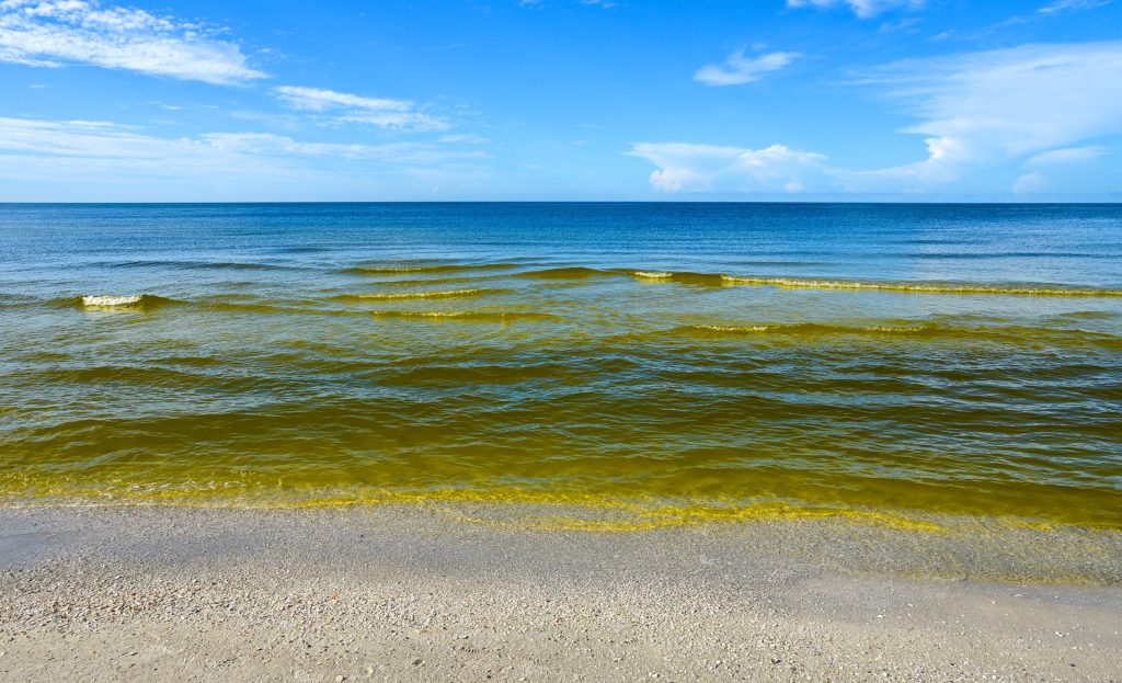
Fig 4. A harmful algal bloom (HAB) in the Gulf of Mexico.
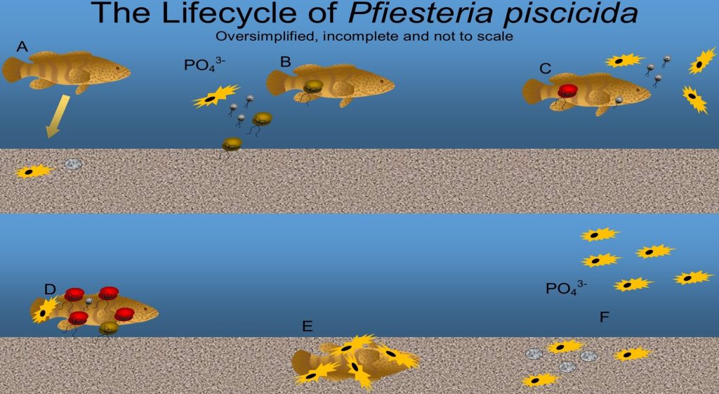
Fig 5. A not to scale oversimplified and incomplete representation of the life cycle of Pfiesteria piscicida. [A] the presence of fish stimulates the germination of cysts. [B] nontoxic zoospores (brown) swarm onto, attach, and begin to feed upon fish. Enriched DIP stimulates the production of large lobose toxic amoeba (yellow), and gametes (grey) [C], whilst zoospores become toxic (red). Fish skin becomes necrotic which reinforces chemical cues, where suspended erythrocytes and more phosphorus hasten gamete production and attacks from large lobose amoebae. [D] overwhelmed narcotized moribund fish descend whilst being eaten alive. [E] carcasses are necrotized by toxin, ingested by amoebae, and biomineralised by microorganisms. [F] elevated phosphorus sustains planktivorous amoebae when fish are lacking, whilst others lay dormant on substrate with cysts. Adapted from Burkholder and Glasgow 1997a, Burkholder and Glasgow 1997, and Litaker and allies 2002. Anticipate commonality with the life cycles and stage transformations of Luciella masanensis, Pfiesteria shumwayae, and Stoeckeria algicida (Jeong et al. 2007a; Jeong et al. 2007). The not illustrated intermediate stages include amoeboid gametes, anisogamous gametes, star-like rhizopodial amoebae, small amoebae, star amoebae, toxic planozygotes, and amoeboid and other forms of cyst (Burkholder & Glasgow 1997a).
A much later study investigated the putative Pfiesteria outbreaks from Chesapeake Bay whose findings proposed that the fish were exposed to Pfiesteria toxins after lesion initiation by Aphanomyces invadans, insofar as wounding would strengthen chemical cues and incite Pfiesteria attacks (Blazer et al. 2016). Exposing teleosts to zoospores of A. invadans resulted in significant mortalities; however, greater incidences arose after subjecting fish to physical trauma (Kiryu et al. 2002).

Fig 6. A scanning electron micrograph (SEM) of a typical dinospore. Image courtesy of The Florida Fish and Wildlife Commission©.
Strains of A. invadans instigate pathogenic infections mostly in estuarine waters, because nil growth was observed on agar supplemented with 12 o/oo sodium chloride (NaCl) while nonpermissive temperatures range from 31 to 35oC (88 to 95oF; Oidtmann 2012). Current knowledge affirms that A. invadans cannot grow at seawater salinities (35 o/oo; ~1.026 SG; Blazer, personal communication) and thus remain no threat to tropical marine ornamentals (Fig 7.).
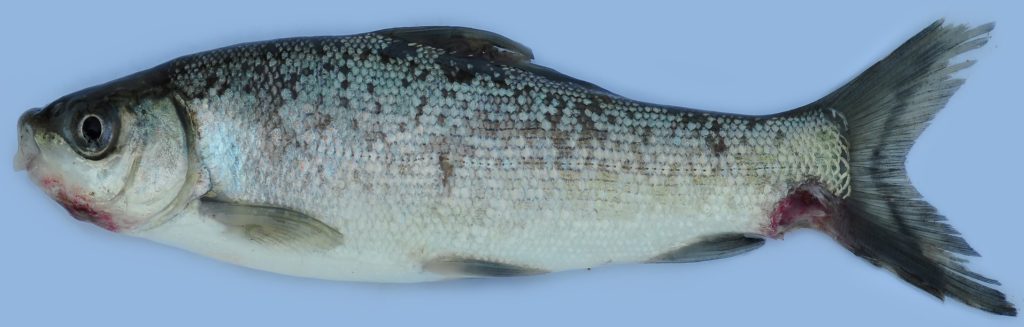
Fig 7. A post-mortem example of freshwater kuria labeo (Labeo gonius) exhibiting epizootic ulcerative syndrome (EUS) caused by the fungal-like chromist Aphanomyces invadans, which is a disease exemplifying hyphae-derived host-specific aetiologies, site-specific ulcerations, petechial hemorrhages, and tail erosion.
Pathogenic “Algae”: Control
Rigorous diagnoses are central to all successful remediations. Pfiesteria piscicida are sustained by plankton when fish are absent, yet their dinospores are bioremediated by sizeable zooplankton which are killed by therapeutic copper (Mallin et al. 1995). Copper sulphate controls toxic cyanobacteria in fish-only systems (Roberts 1989a); however, dinoflagellates exploit purging mechanisms and thus survive therapeutic concentrations (Lage et al. 1996). ~0.05 mg l-1 cupric ions (Cu2+) inhibit the growth of toxic diatoms whereas 96 hours of 0.139 mg l-1 Cu2+ induce their mortality (Lelong et al. 2012a). Nevertheless, 0.1 mg l-1 Cu2+ is the absolute maximum tolerated by marine ornamental teleosts, while ionic copper is toxic to invertebrates including corals and must never be used in reefs.
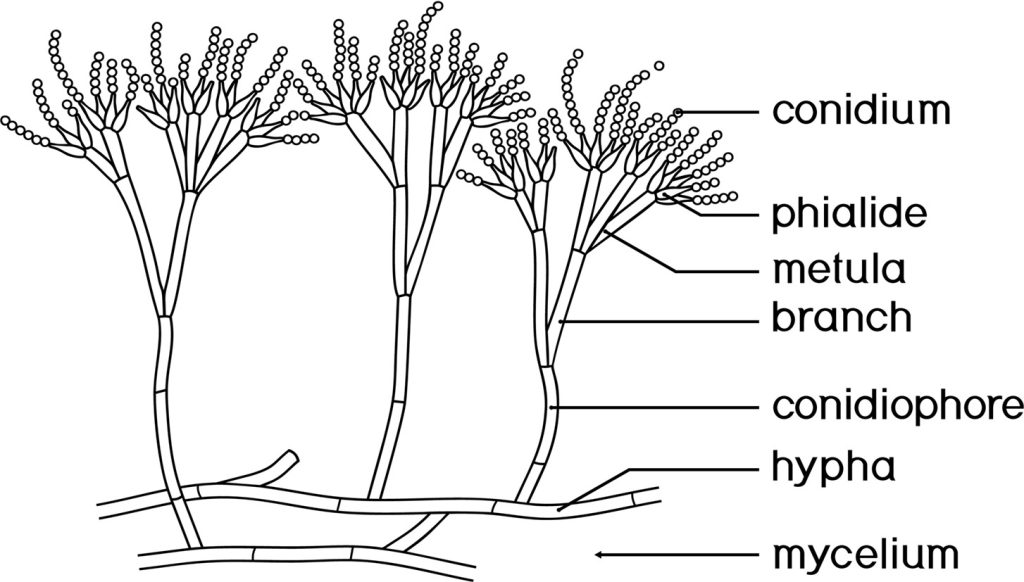
Fig 8. A diagram of fungal anatomy which introduces some unfamiliar terms.
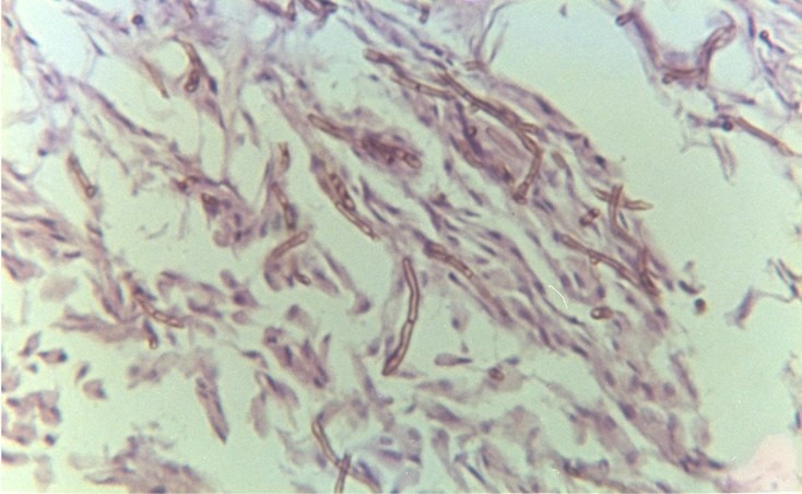
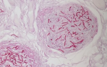


Fig 9. Fungal hyphae instigating necrosis and a profound inflammatory response in the tissues of Atlantic cod (Gadus morhua). Top right – necrotized granulomas. Contains public sector information licensed under the Open Government License (OGL) v3.0. Consult bibliography.
Nevertheless, finfish kill-or-cure is determined by the rate at which they are exposed, and thus gradually increase concentrations over several days. Consult: The Complete Reef Aquarist for vital guidance (https://www.ebay.co.uk/itm/116390701502) or the Reef RanchTM videos available at https://www.reefranch.co.uk/
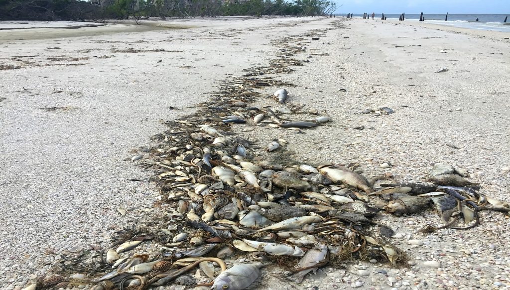
Fig 10. Red tide-induced teleostean mortalities at Fort Myers Beach in Florida.
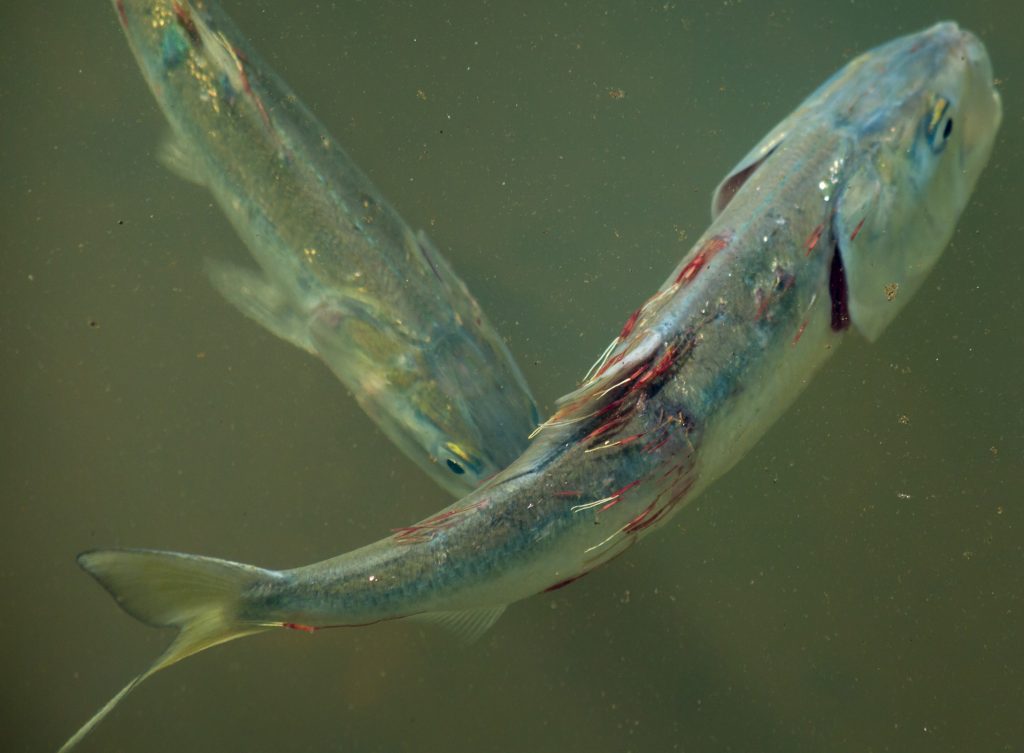
Fig 11. Moribund Atlantic menhaden (Brevoortia tyrannus) heavily infested with marine parasitic copepods causing muscle atrophy (cachexia), dehydration, respiratory and osmotic distress.
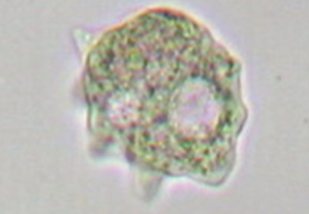
Fig 12. A micrograph of an amoebic gill disease (AGD)-instigating amoeba courtesy of FishPathogens.net.
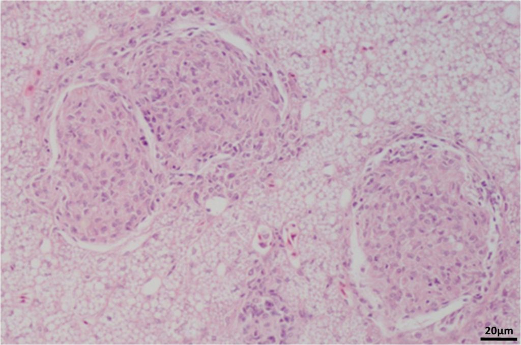
Fig 13. A micrograph of a histologically mounted 5 µm cross-section of granulomas stained with haematoxylin and eosin that appeared as macroscopic nodules in the viscera of Atlantic cod (Gadus morhua). Formed in response to gram negative bacteria of the genus Francisella. Contains public sector information licensed under the Open Government License (OGL) v3.0. Consult bibliography.
Bentonite clay is efficacious which removes both “algae” and toxins from freshwater (Lewis et al. 2003; Sengco et al. 2005; Louzao et al. 2015) which is used in koi ponds but DO concentrations must be considered because saltwater can dissolve merely half as much. Bentonite is therefore not recommended here.
30oC curtails the production of brevetoxin in Heterosigma akashiwo (Heterokonta: Raphidophyceae; Ono et al. 2000), hence rapid increments in heat may prove ameliorating, yet they will be somewhat stressful and may affect the biofilter consortium. Optimized tropical marine ornamental fish-only systems have been run at such temperatures with no observed adverse effect (NOAE). Many dinoflagellates favor hyposalinity and thus maintain 35 o/oo (~1.026 SG; Haque & Onoue 2005) while granular activated carbon (GAC) removes suspended “algae” and sizeable molecular toxins within a matter of hours, but it cannot be used for more than two days and must be prewashed in copious amounts of R/O water. Besides, it removes chemotherapeutics which are essential for commercial teleostean ornamental mariculture, so preclude new specimens and reintroduce therapies over several days.
4 to 12 mg l-1 of formalin proved toxic to red tide microorganisms within 24 hours (Xu et al. 2005); however, research affirms sustained maximum fish-safe exposure is in the region of 22 to 67 times less at 0.18 mg l-1 (ppm; Reardon & Harrell 1990; Francis-Floyd 1996). Exercise care when administering formalin as species sensitivity varies, while it proves harmful to nitrite oxidizers (Keck & Blanc 2002). Moreover, it exerts significant ecological impacts and must never be used in reefs, and there are several precautions one must take before medicating, so it remains unendorsed. Watch: Reef RanchTM videos at https://www.reefranch.co.uk/
Commercial fish-only systems sustain phytoplankton-nurturing concentrations of dissolved inorganic nitrogen (DIN) and DIP and HABs are associated with eutrophy, and thus infections may devastate profitable enterprises, so instigate the cross-contamination avoidance protocols described in Francis-Floyd and Floyd published in 2011.
There are safe ways of lessening system eutrophicants to limit HABs. Kent Marine phosphate sponge is 100 percent safe, mitigates elevated phosphate overnight, and is eminently rechargeable, as opposed to lanthanum chloride whose toxicity has been well documented (Eddy & Bath 1979; Bogers 1995; Mácová et al. 2010). However, nutrient limitation tends to stimulate toxigenesis and mass “algal”-die-off, whereupon a microbial bloom attempts to degrade their vestiges which initiates livestock-decimating hypoxia. Nevertheless, ozone may aid system recovery yet will likely prove insufficient to prevent widespread mortalities. Protracted nutrient downturns and temperature adjustments are advised for chronic to mild infections, whereas “belt and braces” exploits two days of GAC followed by other means of diminishing phosphate to growth-limiting concentrations. It is unwise to apply ozone in the absence of vital insight so review Aslett 2024.

Ultraviolet (UV) sterilization is somewhat ineffective (Frerichs et al. 2000; Bomo et al. 2003; Feldman et al. 2011) yet it may photo-inactivate some toxins like those liberated by the golden alga, Prymnesium parvum/P. parvum f. patelliferum (Barkoh et al. 2010). Superoxide anions (O2–) liberated by H. akashiwo (Kempton et al. 2008; Martnez et al. 2010), spontaneously decompose to hydrogen peroxide (H2O2; de Grey 2002) which improves the antimicrobial efficacy of these peripherals, while sodium thiosulphate (Na2S2O3; tap safe) helps degrade these and other reactive oxygen species (ROS; de Grey 2002; Jorquera et al. 2002; Gonçalves & Gagnon 2018).
Toxic and nuisance dinoflagellates of the genus Ostreopsis form resting cysts at diminished temperatures which remain in stasis at 21oC (70oF) yet germinate at 25oC (77oF; Accoroni & Totti 2016). Reefs benefit greatly from year-round temperatures of 23.5oC (~73.4oF). Lower temperatures increase the growth rates of stony corals (Ruiz-Diaz et al. 2022, cited in Toledo-Hernandez et al. 2023) and suppress the vast majority of coral diseases (Kuta & Richardson 1996; Voss & Richardson 2006; Vega Thurber et al. 2014), while they bolster DO which inhibits red and green slime algae (cyanobacteria).
Most phytoplanktonic blooms common to eutrophic fish-only systems are harmless so verify with brightfield microscopy and leave well alone. Consult: The Complete Reef Aquarist for further guidance now available worldwide (https://www.ebay.co.uk/itm/116390701502).
To learn more, visit the Reef Ranch website.
References
Accoroni, S. & Totti, C. (2016) The toxic benthic dinoflagellates of the genus Ostreopsis in temperate areas: a review. Advances in Oceanography and Limnology. 7(1), 1-15.
Alavi, M., Miller, T., Erlandson, K., Schneider, R. & Belas, R. (2001) Bacterial community associated with Pfiesteria ‐like dinoflagellate cultures. Environmental Microbiology. 3(6),.
Aslett, C., G. (2024) Reef Digress: Fundamental and Foundational Science: Not Simply Water. https://wwww.reefranch.co.uk/
Barkoh, A., Smith, D. & Southard, G. (2010) Prymnesium parvum Control Treatments For Fish Hatcheries. Journal of the American Water Resources Association, 46(1),.
Blazer, V., Phillips, S. & Pendleton, E. (2016) Fish Health, Fungal Infections, and Pfiesteria: The Role of the U.S. Geological Survey. U.S. Geological Survey Fact Sheet. pp 114-198. https://pubs.usgs.gov/fs/1998/114/ [last accessed December 2020].
Blazer, V., S., Densmore, C., L., Lilley, J., H., May, E., B., Vogelbein, W., K. & Zwerner, D., E. (1999) Aphanomycesas a Cause of Ulcerative Skin Lesions of Menhaden from Chesapeake Bay Tributaries. Journal of Aquatic Animal Health. 11(4) 340-349.
Blazer, V., S., Lilley, J., H., Schill, W., B., Kiryu, J., Densmore, C., L., Panyawachira, V. & Chinabut, S. (2002) Aphanomyces invadans in Atlantic Menhaden along the East Coast of the United States. Journal of Aquatic Animal Health, 14(1), 1-10.
Bogers, M. (1995) Daphnia Magna, Reproduction Test With Lanthanum (La), Report No.: 139499 Testing Laboratory: NOTOX BV, Owner Company. https://echa.europa.eu/registration-dossier/-/registered-dossier/14555/6/2/3 [last accessed 27 May 2019].
Bomo, A., Husby, A., Stevik, T. & Hanssen, J. (2003) Removal of fish pathogenic bacteria in biological sand filters. Water Research. 37(11), 2618-2626.
Burkholder, J. & Glasgow, H. (1997) Pjesteria piscicida and other Pfesteria-like dinoflagellates: Behavior, impacts, and environmental controls. Limnology and Oceanography. 42, 1052-1075.
Burkholder, J. & Glasgow, H. (2002) The life cycle and toxicity of pfiesteria piscicida revisited. Journal of Phycology. 38(6),.
Burkholder, J., Glasgow, H., Deamer-Melia, N., Springer, J., Parrow, M., Zhang, C. & Cancellieri, P. (2001b) Species of the toxic Pfiesteria complex, and the importance of functional type in data interpretation. Environmental health perspectives. 109(5), 667-679.
Burkholder, J., Gordon, A., Moeller, P., Law, J., Coyne, K., Lewitus, A., Ramsdell, J., Marshall, H., Deamer, N., Cary, S., Kempton, J., Morton, S. & Rublee, P. (2005) Demonstration of toxicity to fish and to mammalian cells by Pfiesteria species: Comparison of assay methods and strains. Proceedings of the National Academy of Sciences. 102(9), 3471.
Burkholder, J., M. & Glasgow, H., B. (1997a) Trophic Controls on Stage Transformations of a Toxic Ambush‐Predator Dinoflagellate. Journal of Eukaryotic Microbiology. 44, 200-205.
Burkholder, J., M. & Glassgow, H., B., Jr. (1995) Interactions of a Toxic Estuarine Dinoflagellate with Microbial Predators and Prey. Archiv für Protistenkunde. 145(3-4), 177-188.
Burkholder, J., M. (1998) Implications of Harmful Microalgae and Heterotrophic Dinoflagellates in Management of Sustainable Marine Fisheries. Ecological Applications. 8(1), 37-62.
Burkholder, J., M., Glasgow H., B., Jr. & Hobbs, C., W. (1995) Fish kills linked to a toxic ambush-predator dinoflagellate: distribution and environmental conditions. Marine Ecology Progress Series. 124, 43-61.
Burkholder, J., M., Glasgow, H., B., Deamer-Melia, N., J., Springer, J., Parrow, M., W., Zhang, C. & Cancellieri, P., J. (2001) Species of the toxic Pfiesteria complex, and the importance of functional type in data interpretation. Environmental health perspectives. 109(5), 667-679.
Burkholder, J., M., Marshall, H., G., Glasgow, H., B., Seaborn, D., W. & Deamer-Melia, N., J. (2001a) The standardized fish bioassay procedure for detecting and culturing actively toxic Pfiesteria, used by two reference laboratories for atlantic and gulf coast states. Environmental health perspectives. 109(5), 745-756.
de Grey, A., D. (2002) HO2*: the forgotten radical. DNA and cell biology. 21(4), 251-257.
Eddy, F., B. & Bath, R., N. (1979) Effects of lanthanum on sodium and chloride fluxes in the goldfish Carassius auratus. J Comp Physiol B. 129, 145.
Feldman, K., S., Place, A., A., Joshi, S. & White, G. (2011) Water: Baseline Values and Modulation by Carbon Dosing, Protein Skimming, and Granular Activated Carbon Filtration. AdvancedAquarist.com. https://www.advancedaquarist.com/2011/3/aafeature
FishPathogens.net. Images by permission of Dr. Craig Banner (Oregon Department of Fish & Wildlife, retired) and Dr. Stephen Atkinson (Oregon State University).
Francis-Floyd, R. & Floyd. M., R. (2011) Amyloodinium ocellatum, an Important Parasite of Cultured Marine Fish. Southern Regional Aquaculture Center , 1 College of Veterinary Medicine, University of Florida 2 Student, Dept. of Wildlife Ecology and Conservation, University of Florida. SRAC Publication No. 4705.
Francis-Floyd, R. (1996) Use of formalin to control fish parasites. VM-77. College of Veterinary Medicine, Florida Cooperative Extension Service, Institute of Food and Agricultural Sciences, University of Florida, Gainesville, FL. http://edis.ifas.ufl.edu/vm061
Frerichs, G., N, Tweedie, A., Starkey, W., G. & Richards, R., H. (2000) Temperature, pH and electrolyte sensitivity, and heat, UV and disinfectant inactivation of sea bass (Dicentrarchus labrax) neuropathy nodavirus. Aquaculture. 85(1), 13-24.
Glasgow, H., B., Jr., Burkholder, J., M., Schmechel, D., E., Tester, P., A. & Rublee, P., A. (1995) Insidious effects of a toxic estuarine dinoflagellate on fish survival and human health,. Journal of Toxicology and Environmental Health. 46(4), 501-522.
Gonçalves, A., A. & Gagnon, G., A. (2018) Seawater ozonation: effects of seawater parameters on oxidant loading rates, residual toxicity, and total residual oxidants/by-products reduction during storage time. Ozone: Science & Engineering. 40(5), 399-414.
Gottschling, M., Soehner, S., Zinssmeister, C., John, U., Plotner, J., Schweikert, M., Aligizaki, K. & Elbrachter, M. (2012) Delimitation of the Thoracosphaeraceae (Dinophyceae), Including the Calcareous Dinoflagellates, Based on Large Amounts of Ribosomal RNA Sequence Data. Protist. 164(1), 1-15.
Grattan, L., M., Oldach, D., J. & Morris, G. (2001) Human Health Risks of Exposure to Pfiesteria piscicida: Environmental exposure to toxic Pfiesteria piscicida produces a distinct clinical syndrome in some persons; efforts to identify the toxin(s) responsible for the syndrome and to increase understanding of how it disrupts the central nervous system have important implications for public health. BioScience. 51(10), 853-857.
Gwang Hoon, K., Hae Jin, J., Yeong Du, Y., Sunju, K., Ji Hee, H., Jong Won, H. & Giuseppe, C., Z. (2013) Still Acting Green: Continued Expression of Photosynthetic Genes in the Heterotrophic Dinoflagellate Pfiesteria piscicida ( Peridiniales, Alveolata ). PLoS ONE. 8(7), e68232.
Haque, S. & Onoue, Y. (2005) Effects of Salinity on Growth and Toxin Production of a Noxious Phytoflagellate, Heterosigma akashiwo (Raphidophyceae). Botanica Marina. 45(4), 356-363.
Hausmann, K. & Hülsmann, N. (2010) Ultrastructural and functional aspects of static and motile systems in two taxa of the Alveolata: Dinoflagellata and Ciliata. Protistology. 6(3), 139-146.
Jeong, H., J., Ha, J., H., Yoo, Y., D., Park, J., Y., Kim, J., H., Kang, N., S., Kim, T., H., Kim, H., S. & Yih, W., H. (2007) Feeding by the Pfiesteria‐Like Heterotrophic Dinoflagellate Luciella masanensis. Journal of Eukaryotic Microbiology. 54(3), 231-241.
Jeong, H., J., Kim, J., S., Song, J., Y., Kim, J., H., Kim, H., Kim, S. & Kang, N., S. (2007a) Feeding by protists and copepods on the heterotrophic dinoflagellates Pfiesteria piscicida, Stoeckeria algicida, and Luciella masanensis. Marine Ecology-progress Series. 394, 199-211.
Jeong, J., H., Kim, J., S., Kim, J., H., Kim, S., T., Seong, K., Kim, H., Song, J., Y. & Kim, S. (2005) Feeding and grazing impact of the newly described heterotrophic dinoflagellate Stoeckeria algicida on the harmful alga Heterosigma akashiwo. Marine Ecology-progress Series. 295, 69-78.
Jorquera, M., Valencia, G., Eguchi, M., Katayose, M. & Riquelme, C. (2002) Disinfection of seawater for hatchery aquaculture systems using electrolytic water treatment. Aquaculture. 207, 213-224.
Keck, N. & Blanc, G. (2002) Effects of formalin chemotherapeutic treatments on biofilter efficiency in a marine recirculating fish farming system. Aquatic Living Resources. 15(6), 361-370.
Kempton, J., Keppler, C., J., Lewitus, A., Shuler, A. & Wilde, S. (2008) A novel Heterosigma akashiwo (Raphidophyceae) bloom extending from a South Carolina bay to offshore waters. Harmful Algae. 7(2), 236-240.
Kuta, K. & Richardson, L. (2002) Ecological aspects of black band disease of corals: relationships between disease incidence and environmental factors. Coral Reefs. 21, 393.
Kuta, K., G. & Richardson, L., L. (1996) Abundance and distribution of black band disease on coral reefs in the northern Florida keys. Coral Reefs. 15, 219-223.
Lage, O., M., Soares, H., M., V., M., Vasconcelos, M., T., S., D., Parente, A., M. & Salema, R. (1996) Toxicity effects of copper (II) on the marine dinoflagellate Amphidinium carterae: Influence of metal speciation, European Journal of Phycology. 31(4), 341-348.
Lelong, A., Jolley, D., F., Soudant, P. & Hegaret, H. (2012a) Impact of copper exposure on Pseudo-nitzschia spp. physiology and domoic acid production. Aquatic Toxicology. 118-119, 37-47.
Lewis, M., A., Dantin, D., D., Walker, C., C., Kurtz, J., C. & Greene, R., M. (2003) Toxicity of clay flocculation of the toxic dinoflagellate, Karenia brevis, to estuarine invertebrates and fish. Harmful Algae. 2(4), 235-246.
Litaker, R., Vandersea, M., Kibler, S., Madden, V., Noga, E. & Tester, P. (2002) Life Cycle of The Heterotrophic Dinoflagellate Pfiesteria Piscicida (Dinophyceae) 1. Journal of Phycology. 38(3),.
Louzao, M., C., Abal, P., Fernández, D., A., Vieytes, M., R., Legido, J., L., Gómez, C., P., Pais, J. & Botana, L., M. (2015) Study of Adsorption and Flocculation Properties of Natural Clays to Remove Prorocentrum lima. Toxins. 7(10), 3977-3988.
Lun, S., Weidong, L., Jing, W. & Guangjun, S. (2017) Distribution of the toxic dinoflagellate Stoeckeria algicida in Liaodong Bay. Acta Ecologica Sinica. 37(4),.
Mácová, S., Babula, P., Plhalová, L., Žáčková, K., Kizek, R. & Svobodová, Z. (2010) Acute toxicity of lanthanum to fish Danio rerio and Poecilia reticulata. J Biochem Tech. 2(5), 46-47.
Mallin, M., A., Burkholder, J., M., Larsen, M., L. & Glasgow, H., B. (1995) Response of two zooptankton grazers to an ichthyotoxic estuarine dinoflagellate. Journal of Plankton Research. 17(2), 351-363.
Marshall, H., Gordon, A., Seaborn, D., Dyer, B., Dunstan, W. & Seaborn, A. (2000) Comparative culture and toxicity studies between the toxic dinoflagellate Pfiesteria piscicida and a morphologically similar cryptoperidiniopsoid dinoflagellate. Journal of Experimental Marine Biology and Ecology. 255(1), 51-74.
Martnez, R., Orive, E., Laza-Martnez, A. & Seoane, S. (2010) Growth response of six strains of Heterosigma akashiwo to varying temperature, salinity and irradiance conditions. Journal of Plankton Research. 32(4). 529-538.
Moestrup, Ã. & Thomsen, H., A. (1986) Ultrastructure and reconstruction of the flagellar apparatus in Chrysochromulina apheles sp. nov. (Prymnesiophyceae = Haptophyceae). Canadian Journal of Botany. 64(3), 593-610.
Moestrup, Ã., Hansen, G., Daugbjerg, N., Lundholm, N., Overton, J., Vestergard, M., Steenfeldt, S., J., Calado, A., J. & Hansen, P., J. (2014) The dinoflagellates Pfiesteria shumwayae and Luciella masanensis cause fish kills in recirculation fish farms in Denmark. Harmful Algae. 32, 33-39.
Moestrup, Ø. (1994) Economic aspects: ‘blooms’, nuisance species, and toxins. The Haptophyte Algae. Green, J., C. & Leadbeater, B., S., C. (eds.). Systematics Association Special Vol. 51, Clarendon Press, Oxford. pp 265-285.
Noga, E. (2000a) Review Article: Skin Ulcers in Fish: Pfiesteria and Other Etiologies. Toxicologic Pathology. 28(6), 807-823.
Noga, E., J. (1989) Culture conditions affecting the in vitro propagation of Amyloodinium ocellatum. Diseases of Aquatic Organisms. 6, 147-143.
Noga, E., J. (2000) Fish Disease: Diagnosis and Treatment, First Edition. Noga, E., J. (ed.). Ames, IA: Iowa State University Press. US.
Noga, E., Khoo, L., Stevens, J., Fan, Z. & Burkholder, J. (1996) Novel toxic dinoflagellate causes epidemic disease in estuarine fish. Marine Pollution Bulletin. 32(2), 219-224.
Oidtmann, B. (2012) Review of Biological Factors Relevant to Import Risk Assessments for Epizootic Ulcerative Syndrome (Aphanomyces invadans). Transboundary and Emerging Diseases. 59, 26-39.
Ono, K., Khan, S. & Onoue, Y. (2000) Effects of temperature and light intensity on the growth and toxicity of Heterosigma akashiwo (Raphidophyceae). Aquaculture Research. 31(5),.
Open Government Licence (OGL) v3.0. Contains public sector information licensed under the Open Government Licence v3.0. Inclusion of such information does not constitute any official status or UK Government endorsement of the use of the information in this publication.
Reardon, I., S. & Harrell, R., M. (1990) Acute toxicity of formalin and copper sulfate to striped bass fingerlings held in varying salinities. Aquaculture. 87(3), 255-270.
Roberts, R., J. (1989a) Fish Pathology. Second Edition. Roberts. R., J. (ed.). Bailliere Tindall, 24-28 Oval Road, London NW1 7DX, UK. pp 336; pp 302-303.
Ruiz-Diaz, C., P., Toledo-Hernández, C. & Sánchez, J., L. (2022) The effects of depth-related environmental factors and source of collection on life history traits in Acropora cervicornis raised in nurseries. Water J. 14, 212. https://doi.org/10.3390/w14020212
Samet, J., Bignami, G., S., Feldman, R., Hawkins, W., Neff, J. & Smayda, T. (2001) Pfiesteria: review of the science and identification of research gaps. Report for the National Center for Environmental Health, Centers for Disease Control and Prevention. Environmental health perspectives. 109(5), 639-659.
Schnepf, E. & Elbrachter, M. (1992) Nutritional Strategies in Dinoflagellates – A review with emphasis on cell biological. European journal of protistology. 28(1), 3-24.
Schulz, F., Tyml, T., Pizzetti, I., Dyková, I., Fazi, S., Kostka, M. & Horn, M. (2015) Marine amoebae with cytoplasmic and perinuclear symbionts deeply branching in the Gammaproteobacteria. Scientific Reports. 5(1), 1-10.
Sengco, M., Hagstrom, J., Graneli, E. & Anderson, D. (2005) Removal of Prymnesium parvum (Haptophyceae) and Its Toxins Using Clay Minerals. Harmful Algae. 4, 261-274.
Springer, J., J., Shumway, S., Burkholder, J., A., M. & Glasgow, H., B. (2002) Interactions between the toxic estuarine dinoflagellate Pfiesteria piscicida and two species of bivalve molluscs. Marine Ecology-Progress Series. 245, 1-10.
Steidinger, K., Burkholder, J., Glasgow, H., Hobbs, C., Garrett, J., Truby, E., Noga, E. & Smith, S. (1996) Pfiesteria Piscicida Gen. Et Sp. Nov. (Pfiesteriaceae Fam. Nov.), A New Toxic Dinoflagellate With A Complex Life Cycle and Behavior. Journal of Phycology, 32(1),.
Toledo-Hernandez, C., Ruiz-Diaz, C., P., Ramirez-Lugo, J., S., Torres-Diaz, M., Santiago-Pagan, L., Bruno-Chardon, A. & Diaz-Vazquez, L., M. (2023) Uncovering the link between environmental factors and coral immunity: A study of fluorescent protein expression and phenoloxidase activity in Acropora cervicornis. Forntiers in Marine Scinence. https://dio.org/10.3389/fmars.2023.1133486
Vega Thurber, R., L., Burkepile, D., E., Fuchs, C., Shantz, A., A., McMinds, R. & Zaneveld, J., R. (2014) Chronic nutrient enrichment increases prevalence and severity of coral disease and bleaching. Glob Change Biol. 20, 544-554.
Voss, J., & Richardson, L. (2006) Nutrient enrichment enhances black band disease progression in corals. Coral Reefs. 25(4), 569-576.
WoRMS (2022a) Pfiesteriaceae Steidinger & Burkholder, 1996. MarineSpecies.org. https://www.marinespecies.org/aphia.php?p=taxdetails&id=246596
WoRMS (2022b) Thoracosphaeraceae Schiller, 1930 emend. Tangen in Tangen, Brand, Blackwelder & Guillard, 1982. MarineSpecies.org. https://www.marinespecies.org/aphia.php?p=taxdetails&id=109438
WoRMS (2022c) Tubulinea. MarineSpecies.org. https://www.marinespecies.org/aphia.php?p=taxdetails&id=582188
Wu, R., S., S. & Woo, N., Y., S. (1983) Tolerance of hypo-osmotic salinities in thirteen species of adult marine fish: Implications for estuarine fish culture. Aquaculture. 32(1), 175-181.
Xu, H., Song, W., Zhu, M., Qiu, Y. & Ma, H. (2005) Acute toxicity

0 Comments