By Tim Wijgerde
The colorful appearance of marine life is the main reason why many people choose to maintain a salt water aquarium at home. Phenomena known as iridescence and fluorescence result in highly attractive marine fishes, corals and other invertebrates. When we examine these ocean dwellers in detail through a camera lens, a fascinating world is revealed to us, with the most elaborate structures and colors imaginable. This article will provide such a detailed view of marine life using macro and close-up photography.
Also make sure to take a look at another article featured in this issue of Reefs Magazine, “Macro Photography in Coral Reef Aquariums,” by Sanjay Joshi. His very detailed selection introduces the basics of macro photography, as well as a number of advanced techniques, and succinctly explains the benefits behind using various pieces of photographic equipment.
Fishes
It is no secret that marine fishes are amongst the most colorful creatures on earth. Both in the wild and in captivity, they are the first to attract the attention of the casual onlooker. Available in many forms and colors, they provide the aquarium with movement and fascinating behavior. The exquisite colors of fish (and of any other creature for that matter) serve to attract mates, fend off predators and competitors, or provide camouflage.
Highly popular marine fishes are those from the wrasse family (Labridae), which can display striking patterns and colors. Although their fast, erratic movements and sometimes secluded behavior make it challenging to study these fishes in detail, a close-up view reveals a kaleidoscope of colors and patterns. What is interesting about wrasses is that they are sequential hermaphrodites, meaning they can change sex during their life. Most wrasses exhibit a phenomenon known as protogyny (from the Greek words protos, or first, and gunē, or female), where the fishes start their lives as females, and can change to males in an all-female group (thus in the absence of a dominant alpha male). This socially driven sex change is sometimes accompanied by a dramatic change in coloration, and with sufficient knowledge, one can identify the alpha male within a group of wrasses.
Another colorful fish with a secluded lifestyle is Koumansetta rainfordi, formerly known as Amblygobius rainfordi (family Gobiidae). This small fish is often housed in groups and usually hides underneath rocks and within crevices. It displays typical red/orange bands, which run lengthwise along the body and extend from the base of the tail to the mouth. A camera flash reveals a green to dark grey epithelium (skin) that serves as a background for deep red bands.
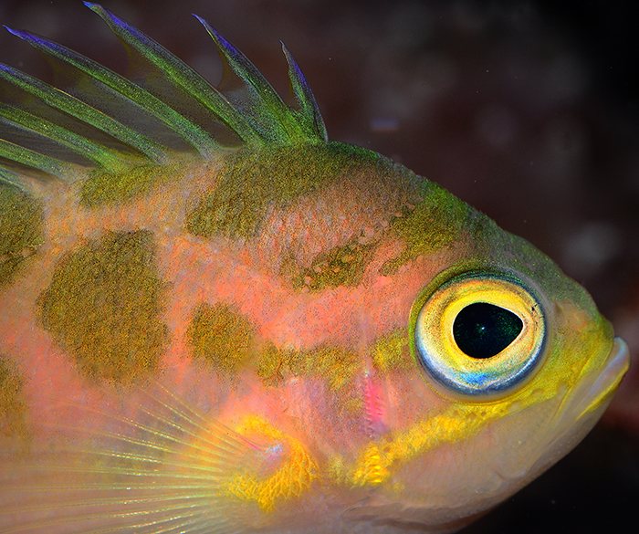
In terms of iridescence, Odontanthias borbonius (family Serranidae) is a real winner. This species is found across the Indo-Pacific, and has become increasingly popular as an aquarium fish over the last few years. The sides and dorsal spines of this species display remarkable iridescence, with blue, green and yellow as the dominant colors. As O. borbonius usually inhabits deep waters, at an approximate depth range of 70 to 300 meters (or 233 to 1000 feet), its colors can only be observed in the wild using a strobe light or camera flash.
Scleractinian Corals
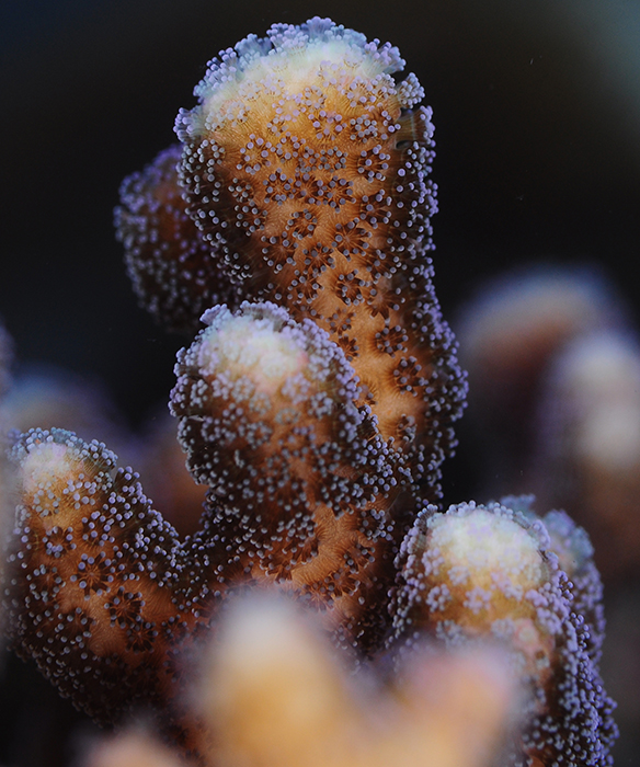
Amongst the most popular invertebrate animals in the aquarium hobby today are the stony or scleractinian corals. With their vibrant colors and diverse shapes, these often static animals are highly suitable models for close-up and macro photography. So too the species Stylophora pistillata (family Pocilloporidae), with its small polyps that come in shades of purple, pink, green and beige. These colors are matched with a brownish coenenchyme, a result of the symbiotic dinoflagellates (zooxanthellae) which occupy the coral’s tissue. S. pistillata is true lab rat, widely used in scientific studies, but it’s also a hardy and attractive species that grows well in the average aquarium.
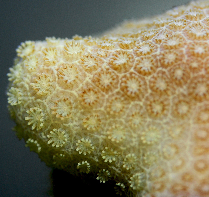
Another small polyped scleractinian coral that catches the eye is Porites cylindrica, an Indo-Pacific coral with a conspicuous banana-yellow coloration. Although its polyps are very small, a close-up view reveals that each one bears twelve tentacles, just like those of S. pistillata. In fact, all scleractinian corals exhibit a multiple of six tentacles, and thus have hexaradial (six-fold) symmetry. This is why stony corals are members of the subclass Hexacorallia.
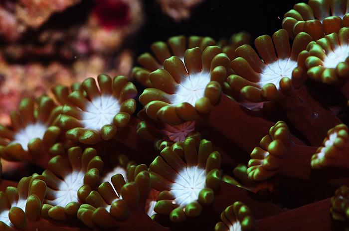
A scleractinian coral with much larger polyps is Alveopora gigas (family Poritidae). This delicate species has polyps with very long stems, which can extend over two inches from the corallites (depressions in the coral skeleton which house the individual polyps). Although it is related to P. cylindrica, A. gigas has a very different appearance, with long polyps that gently wave about in the water current.
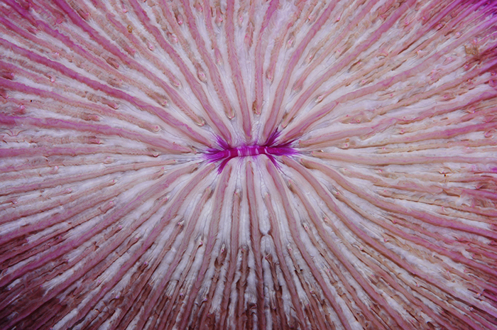
In terms of large polyps, corals from the Fungiidae family take the cake. These corals are free-living, and often consist of just a single polyp with a central mouth. Fungiid corals have polyps that can attain diameters of over 10 inches across, and their large mouths allow one to study their gluttonous feeding behavior with the naked eye.
Octocorals
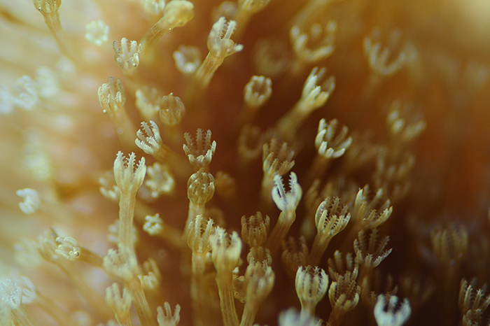
Although not so popular as in the 1980’s and 90’s, octocorals are found in most saltwater aquaria. In contrast to stony corals with hexaradial symmetry, all octocorals—as their name suggests—have octoradial or eight-fold symmetry, with eight tentacles on each polyp. Many species belonging to this subclass only show modest coloration. An example are corals from the genus Sarcophyton, which usually are different shades of yellow or brown. When we zoom in on these animals, however, Sarcophyton spp. are just as striking in their appearance as any other coral. They actually have two types of polyps; the autozooids, which serve as feeding and reproductive units, and smaller, inconspicuous siphonozooids, which take care of water transport and hydrostatic pressure of the colony.
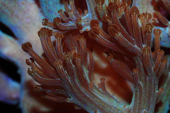
Some octocorals that seem to have little pigmentation appear to hide their spectacular colors. Although most specimens within the genus Heteroxenia are shades of brown or grey, some possess a blue-greenish hue. Although by itself this is attractive enough, a camera flash reveals a hidden speckled pattern with many colors, reminiscent of fireworks in a night sky. Their pulsating polyps further add to the beauty of these corals.
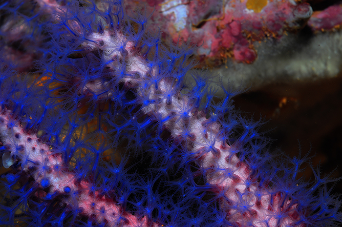
Corals which require no artificial flash to reveal their true splendor are the gorgonians (family Gorgoniidae). A nice example are Guaiagorgia spp., which have deep blue polyps. The tissue that connects the individual polyps, the coenenchyme, is slightly pink, resulting in a beautiful contrast. Upon close inspection, these gorgonian polyps have tentacles that each bear tiny protrusions called pinnules. These increase the coral’s surface area to enhance prey capture and gas exchange. Their feeding behavior can be recorded with a camera and macro lens, which reveals how individual tentacles quickly wrap themselves around prey items, one by one.
Corallimorpharians
The corallimorpharians are often a subject of confusion amongst hobbyists. With their soft, flattened bodies, they resemble anemones more than true corals. However, these creatures are actually grouped within the separate order Corallimorpharia, as relatives of stony corals (order Scleractinia) and true anemones (order Actiniaria), amongst others. Recent genetic evidence suggests that corallimorpharians are strongly related to some scleractinian corals, and that they lost their ability to produce a skeleton during the Cretaceous, when oceans were more acidic.

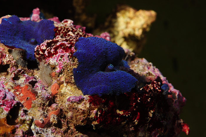
Ricordea florida. Note the flatworms (possibly Waminoa sp.) on the upper and lower sides of the oral disks. Photos by Tim Wijgerde.
Species from the genera Discosoma, Ricordea and Rhodactis are popular in the aquarium trade, with highly colorful and intricately patterned specimens available.Ricordea florida is a species with a peculiar morphology, having bulbous (papillae) structures on its oral disk. These papillae are actually tentacles, and they are usually colored in a circular pattern on the oral disk, for example green in the peripheral part of the disk, and orange in the central part. The tissue in between of the papillae can have different colors, such as pink, purple or blue.
Discosoma spp. exhibit attractive striped and dotted color patterns, and wart-like tentacles called verrucae. Sometimes they are uniform in coloration, and deep blue specimens seem to be the most sought-after. These easy to care for creatures do well in moderately lit environments, and often encrust the darker areas of coral reefs and aquaria.
Zoanthid Corals
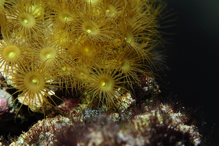
Zoanthid corals are also members of the subclass Hexacorallia, just like stony corals, anemones and corallimorpharians, and are grouped within the order Zoantharia (formerly called Zoanthidea). Zoanthids are colonial animals, with the genera Palythoa, Zoanthus and Parazoanthus often found in home aquaria, including the well-known yellow Bali polyps (Parazoanthus sp.). This popularParazoanthus sp. has polyps bearing 24 tentacle pairs, with a grand total of 48 tentacles. This number is not trivial, as this multiple of six again reveals six-fold symmetry, which is why Parazoanthus spp. are members of the Hexacorallia subclass.
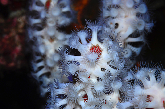
A tentatively identified Parazoanthus sp., symbiotically growing on the sponge Trikentrion flabelliforme. Photo by Tim Wijgerde.
Although many zoanthids live as separate colonies, several species have formed an interesting symbiosis with sponges. This possibly includes a white Parazoanthus sp., which is found together with the red sponge Trikentrion flabelliforme. This zoanthid-bearing sponge is sometimes referred to under dubious names, such as Spider Sponge and White Line Sponge. Found in the Arafura Sea, north of Australia, T. flabelliforme forms a beautiful red contrast with its stark white zoanthid symbiont. The zoanthid polyps grow on the sponge’s pinacoderm, or skin, making this symbiosis potentially superficial. Some scientific evidence suggests that this symbiosis is an example of parasitism, as the zoanthids seem to reduce the sponge’s filtering capacity. Unfortunately, T. flabelliforme usually does not thrive in heavily filtered aquaria, as it requires fine particulate and dissolved matter to feed on. Its zoanthid symbiont also requires zooplankton to grow, as it does not host zooxanthellae and cannot make use of light for its sustenance.
Clams
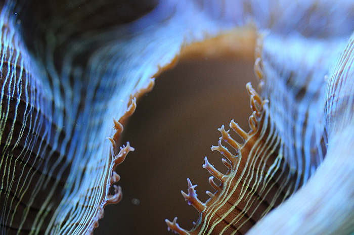
Clams (phylum Mollusca, class Bivalvia) are fascinating mollusks that appear in all shapes and sizes, across all oceans. Species of main interest to aquarists are those from the genus Tridacna, which are host to symbiotic dinoflagellates (or zooxanthellae). The clams feed on the nutrients produced by their symbiotic zooanthellae, which utilize (artificial) light to convert carbon dioxide to glycerol, glucose and other organic compounds. In addition, these clams use their filtration apparatus to remove particles from the water, including algae, which serve as an additional food source. Water is pumped through the incurrent siphon, after which particles are filtered by the clam’s gills and transferred to the mouth using mucus. Finally the filtered water leaves the animal through the excurrent siphon. Upon close inspection, tentacles lining the edge of the incurrent siphon can be seen on specific clams. An example is Tridacna derasa, which has branched tentacles on the edges on its incurrent siphon. It has been suggested that these function as a prefilter, to prevent large particles—which may cause harmful obstructions—from entering the siphon (Fishelson 2000).
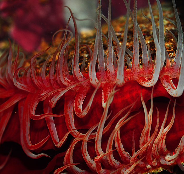
A fascinating mollusc which is regularly found in the aquarium trade is Ctenoides scabra (having many old, now invalid synonyms, including Lima scabra). This Caribbean clam is popularly referred to under the name flame scallop, as its fleshy mantle appears to be literally on fire. This is due to a high concentration of caroteinods in the tissue of these clams (Lin and Pompa 1977). To add further spectacle to the blazing red color of file clams, the Indo-Pacific species C. alesseems to produce its own lightning! This attractive phenomenon, known as bioluminescence, is one of the reasons why they are maintained in captivity. Unfortunately, they usually do not live long in aquaria, as phytoplankton is not available to them in sufficient quantities.
Polychaetes
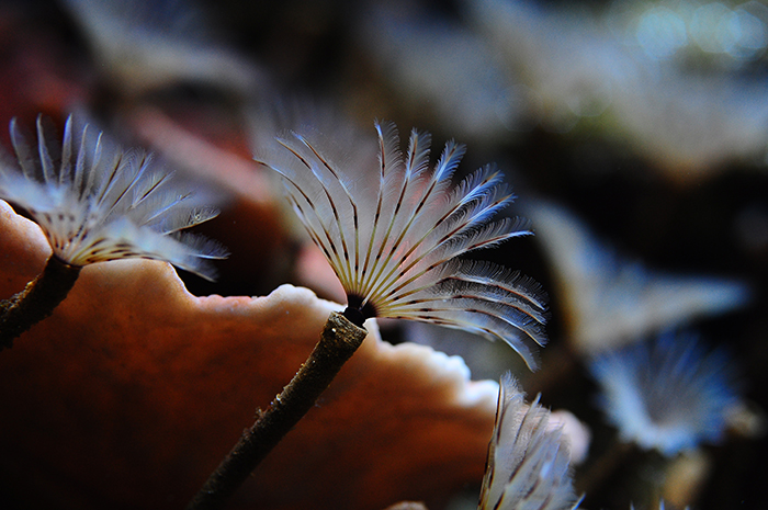
Sabellid polychaete, with two clusters of radioles on its head (prostomium) that create a fan-like structure. Photo by Tim Wijgerde.
Polychaetes are amongst the less spectacularly colored marine animals, and they generally live a secluded lifestyle in between rocks. Polychaetes that are highly common on coral reefs, intertidal zones and in aquaria are the so-called feather duster worms. These are polychaetes from the Sabellidae family, and they have a head segment covered in delicately branched, feathery structures called branchiae. These branchiae, or gills, are used for respiration (breathing) and food capture. They consist of two fan-shaped clusters that are each made up of feathery tentacles, or radioles. Each radiole has fine pinnules, or side branches, allowing sabellid polychaetes to capture very small plankton including bacteria, protists and algae. The radioles each have a food groove covered in cilia, microscopic hairs that transport food particles to the worm’s mouth using mucus. This process is highly similar to the feeding mechanism of crinoids. The filter feeding behavior of sabellid polychaetes is why these worms do best in particle-rich aquaria. They are often found in filtration sumps and overflow chambers, where fine detritus may allow them to thrive. They build their parchment-like tubes by secreting certain proteins, and will fully retract in their tube when disturbed.
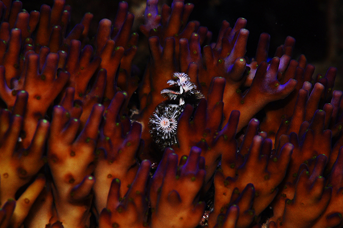
Serpulid polychaete growing in an Acropora colony, with typical radioles that form two spiral patterns, akin to Christmas trees. Photo by Tim Wijgerde.
In terms of modest coloration, Spirobranchus spp. (family Serpulidae) are an exception to the rule. These polychaetes are commonly referred to as Christmas tree worms, and they are found in many colors, including combinations of yellow, red and blue. Spirobranchus spp. are usually found growing in Porites corals. Unlike the Sabellidae, these worms possess an operculum, a modified radiole that allows the worms to close off their tubes after retraction. In addition, these serpulid worms secrete a tube made of calcium carbonate, which is a lot tougher than the parchment-like ones produced by sabellids.
Ascidians
Ascidians belong to the more obscure animals found in the marine aquarium. Their cryptic lifestyle, together with their usually small and translucent bodies, causes them to be often overlooked. Ascidians are true filter feeders, able to filter many gallons of water per day, removing bacteria and other particles from the water. To this end, water is drawn into the branchial/buccal siphon by ciliated cells of the pharynx (throat), which create water movement by simultaneously beating their hair-like cilia. The pharynx is covered by a layer of mucus, to which food particles stick. This mucus is secreted by gland cells of the endostyle, a ciliated groove located closely to the pharynx. After capture, food particles are transported to the oesophagus together with mucus, and digested and absorbed in the stomach and intestinal tract, respectively. After the water has been filtered in the pharynx, it eventually leaves the animal through the atrial, or excurrent siphon. The siphons allow tunicates to deflate by pumping water from their body cavity. During this process, the buccal siphon is closed off. They inflate by opening up their buccal siphon, and closing off the atrial siphon.
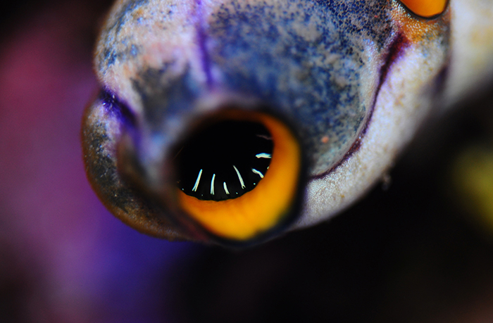
Although ascidians are often overlooked, some species can be both large and colorful. So too the more familiar species called Polycarpa aurata, which is regularly sold in the aquarium trade. Its purplish tunic, or mantle, is often speckled with yellow and blue dots which makes it an attractive animal. In addition, its incurrent and excurrent siphons have orange rings on their inner sides, making the animal highly colorful. Upon close inspection, the incurrent, buccal siphon also has white tentacles, which may prevent large particles from being drawn into the animal, in a similar way to the branched tentacles on Tridacna siphons.
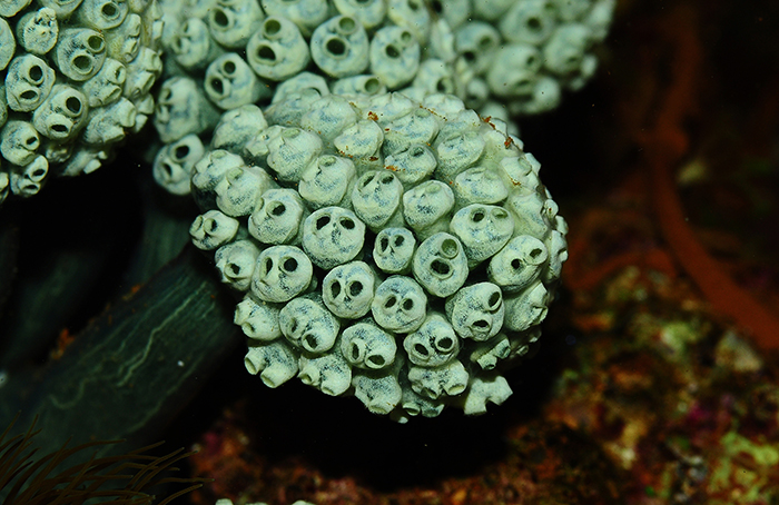
Other species can be colonial, and share various body parts. A nice example is the species Neptheis fascicularis, with the individual zooids clustered in groups, making the entire colony somewhat resemble certain fungi. Although ascidians are interesting and colorful animals, they are difficult to maintain in captivity due to their dependence on small food particles, just like the sponges, zoanthids and clams discussed above.
Concluding Remarks
It seems that the wonderful world of marine life, with its complex features and colors, is best observed through a camera lens. Macro and close-up photography provide a detailed view of sea creatures, teaching us about their form and function. This truly heightens our appreciation for them, I think.
References
Lin AL, Pompa LA (1977) Carotenoids of the red clam Lima scabra. Bol Inst Oceanogr Univ Oriente 16:83–86.
Fishelson L (2000) Comparative morphology and cytology of siphons and siphonal sensory organs in selected bivalve molluscs. Mar Biol 137:497–509.




0 Comments