

When I wrote Aquarium Corals, I was beginning to read some papers that suggested that stony corals, like soft corals, might produce their fair share of secondary metabolites. By this, I mean that while early investigators either failed to look for – or failed to find – bioactive chemicals in stony corals, some recent investigations indicated that they are indeed present. It had been largely assumed that, because stony corals had a protective skeleton, that they had little need for protective chemicals. In contrast, soft corals, sponges, and other soft bodied invertebrates were studied extensively and found, as would be expected of sessile animals, that a copeia of bioactive compounds were produced and stored or released by them for a multiplicity of roles. Among these were allelopathic chemicals used in defense, space competition, and as anti-predation substances. Additional roles were discovered including roles in protecting eggs, cueing settlement, attracting sperm, and others. As massive numbers of novel compounds were found in these taxa, stony corals remained largely ignored.
I was able to find some direct and indirect references to the production of these metabolites in stony corals, and one of the most direct involved a “toxic exudate” from Goniopora tenuidens (Gunthorpe and Cameron 1990a). Whether forgetting to retrieve them, or not noticing the other references in that paper, I wrongfully assumed this was all that had been written on the subject. It was not until doing a literature search for the toxic effects of metals on stony corals for a discussion between Ron Shimek, Randy Holmes- Farley, and others (http://www.reefcentral.com/vbulletin/showthread.php?s=ef6 e4e6f80f5ab895950901faef0ed84&threadid=100591) that I stumbled across another paper. This time, I was observant enough to notice that not a few, but many other articles had been written on the subject. Interestingly, and perhaps among the reasons I had not found these papers before, was that the majority of them were to be found in the medical literature. Soon, I found myself completely stunned at what I had learned, for little of this is new to science. It is however, new to me, and I believe will be new to most.
In the Gunthorpe and Cameron (1990a) paper, it was found that a “range of bioactivity, as aqueous extracts from Australian specimens displayed consistent toxicity to mice and cytolytic activity, while exhibiting interspecific variation in antibiotic activity and ichthyotoxicity.” An investigation was done to determine the effect of alellochemicals previously described (Gunthorpe and Cameron 1990b) as being toxic to scleractinians; in this study, the effects of G. tenuidens towards Galaxea fascicularis. Individual polyps of Galaxea were placed in aerated seawater with 3-4 cm portions of G. tenuidens and examined after 0, 12, and 24 hours (called “conditioned seawater). They were then transferred to another tank with flowing seawater and observed. “Conditioned seawater was determined to be toxic to a test species when all corals of that species showed signs of intoxication.” It was found that conditioned seawater of 8 of 10 Goniopora colonies was toxic to at least one of the test species, and the signs of intoxication were consistent in all cases. Goniopora exudate was lethal to Galaxea fascicularis, inducing sustained polyp contraction, increased mucus production, loss of tissue coloration, and loss of tissue from the skeletal matrix. No Goniopora exudate was lethal to other Goniopora. Sub- lethal signs included polyp contraction and increased mucus production reversed after a half hour exposure to fresh seawater. The fact that Goniopora species could cause such a reaction and death in the strongly aggressive Galaxea within one day is remarkable. It is, however, only the beginning.
Gunthorpe and Cameron (1990c) have yet another study using_ Lobophyllia coymbosa, Favites abdita, Favia matthaii, Favia stelligera, Platygyra daedaelea, Leptoria phrygia, Cyphastrea serailia, Hydnophora exesa, and Astreopora myriophthalma as test subjects._ In this study, they examined toxicity to mice, toxicity to a coral (_Galaxea_ fascicularis), toxicity to a hydroid (_Solandaria fusca_), cytolytic activity on sheep erythrocytes and sea urchin ova, and for antibacterial activity against eight bacterial species. They found that regardless of species, extracts from these corals induced sustained contraction of polyps, increased mucus production, loss of tissue coloration, and loss of tissue from the skeletal matrix. In summary, extracts of each colony of each species produced a positive response in at least two of the assays and activity was highest in the activity against corals and mice, and also in cytolysis of red blood cells.
The next Gunthorpe and Cameron paper (1990b) examined aqueous extracts of 58 scleractinian species from 11 families (Table 1).
| None | None | None |
|---|---|---|
| Favites (5 species) | Favia (7 species) | Cyphastrea (2 species) |
| Platygyra (1 species) | Montastrea (3 species) | Goniastrea (5 species) |
| Leptoria (1 species) | Leptastrea (1 species) | Oulophyllia (1 species) |
| Australogyra (1 species) | Echinopora (1 species) | Porites (1 species) |
| Goniopora (1 species) | Hydnophora (1 species) | Merulina (1 species) |
| Pavona (2 species) | Astreopora (1 species) | Acropora (5 species) |
| Coscinarea (2 species) | Psammacora (2 species) | Galaxea (1 species) |
| Acrhelia (1 species) | Fungia (1 species) | Pocillopora ( 1 species) |
| Seriatopora (1 species) | Turbinaria (1 species) |
This experiment utilized several toxicity assays. In the first, mouse toxicity was determined by effects of coral extracts in producing loss of physical responses, lethargy, reduced body temperature, diarrhea, and death by respiratory paralysis (with the heart still beating post-mortem). They found at least one extract from 93% of the species examined was toxic to at least one test species. Results showed that 38% of 274 colonies assayed were toxic to mice (at least one colony of 41 species). The most toxic was Favites abdita and the least toxic was Leptoria phrygia. The extracts were found to be slow acting toxins, although there was a range; Pavona decussata induced death to mice in 1.3+/- 0.4 hours and Goniopora tenuidens induced death within 45 minutes. Extracts of 71% of the toxic colonies caused death within 24 hours, 28% within 6 hours. The amounts of extract provided by injection to mice were 0.5 ml of an extract prepared from the tissue digest of samples or small colonies in 100 ml of water. For example, a single Fungia would produce enough toxin to kill 200 mice within 24 hours – and Fungia was not nearly the most toxic coral in this study!
The second toxicity assay was done by determining ichthyotoxicity to the mosquitofish, Gambusia affinis, by placing the fish in 70 ml of water with 500 mg/l concentrations of the extracts. Only four species (_Seriatopora hystrix, Acropora cuneata, Goniopora tenuidens_, and Pavona decussata) were toxic to the mosquitofish. Signs of toxicity included hypoactivity, ataxia, and reduced visual response to stimuli.
The third toxicity assay examined the ability of 500 mg/l concentration of coral extract to lyse sheep red blood cells. At least one colony of 49 separate species of 57 tested (153 of 240 colonies, or 64%) was lytic. A majority (73%) were potent lysins, lysing more than 90% of erythrocytes. Favites flexuosa was the most lytic, and Goniastrea australensis was the least lytic.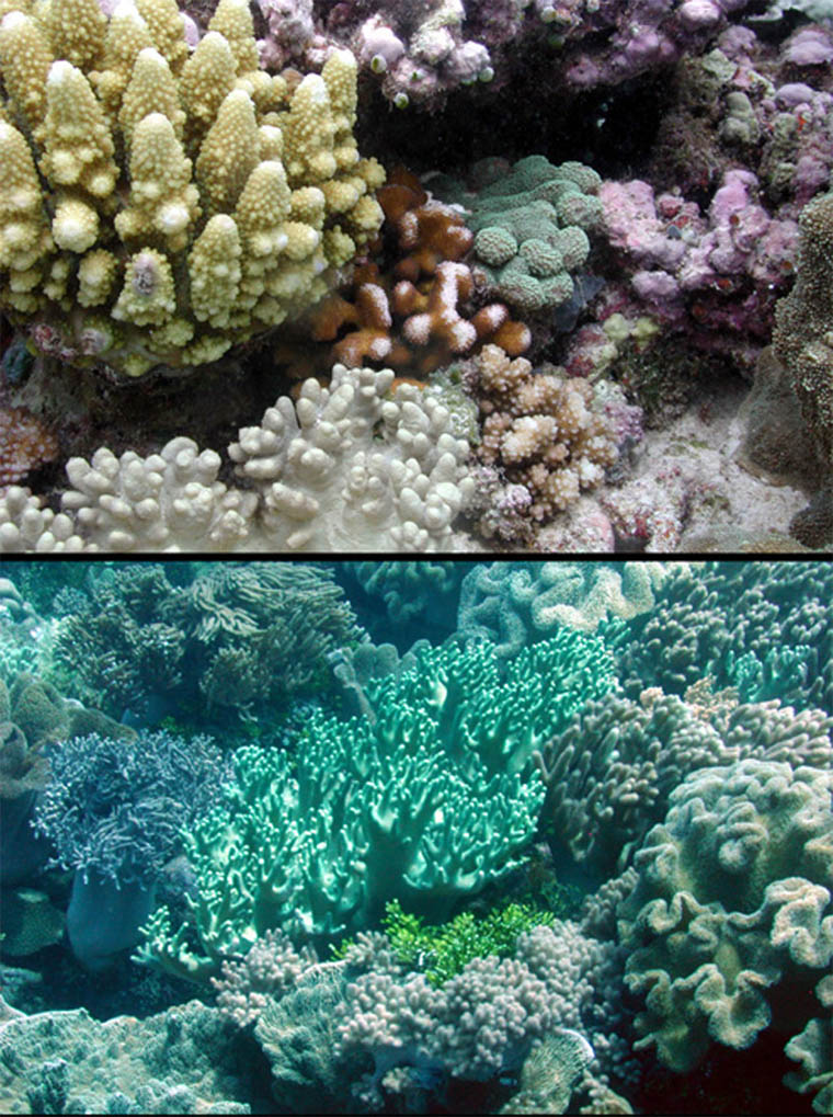
A final antimicrobial activity assay was done, and results were similar to those in Koh (1997a, 1997b, and many others with soft corals and gorgonians), in that most species showed some to considerable activity against bacteria. Only five species were found to be inactive, and this may be due to the types of bacteria used in the tests – that these species may not have activity against those tested but may have antibiotic effects against others as Koh found. In general, Lobophyllia and Symphyllia extracts were highly antibiotic in this study.
It is worth mentioning that nematocysts were considered in this study, and no relationship between discharged nematocysts and incidence or type of activity of the extracts was found. The type of toxicity and activity found here is very similar to the degree, ranges and types of activity found in sponges, soft corals, algae, and other sessile reef invertebrates (McCaffery 1988, Coll et al. 1982, Bakus 1986 and others listed in references). In conclusion, the study found that toxins and cytolysins are widespread in stony corals, although variations in strength exist within and across species (Table 2).
Areas of densely packed stony corals (above) and soft corals (below), sometimes referred to as “coral gardens” are common on coral reefs. However, both communities produce bioactive chemicals that have many effects, including toxic ones. The effect of dilution by sheer ocean volume is probably is a primary reason for the existence of such crowded conditions in the wild. Aquariums, with small closed water volumes, lack this advantage and may suffer from the consequences of high levels of toxins produced by all manner of marine plants and animals, including stony corals. Photos: Eric Borneman
| Assay | number of species | notable |
|---|---|---|
| Toxic to mice | 71% of 58 species | Goniopora and Pavona highly toxic |
| Haemolytic activity | 86% of 57 species | Fungia and Oculinidae not haemolytic |
| Antimicrobial activity | 65% of 55 species | Lobophyllia and Symphyllia highly active |
| Ichthyotoxic activity | 9% of 45 species | |
| Bioactivity, total | 91% of 58 species |
An even earlier study found aqueous extracts from Goniopora gracilis, G. tenuidens, G. planulata, Cyphastrea chalcidicum, Pavona ebtusata and_ an Acropora_ sp. were toxic to mice (Hashimoto and Ashida 1973). A later study by Kaul et al. (1977) found substances pharmacologically active on mammalian cardiovascular, motor, and CNS systems to be produced by Acropora cervicornis, A. paniculata, A. palmata, Fungia fungites, Goniastrea retiformis and_ Montipora marshallensis_. Grozinger (1983) found a biologically active compound in Madracis mirabilis that is also found in nudibranchs and sponges. Stony corals can also inhibit growth of marine algae (De Ruyter van Steveninck 1988). Finally, Sheppard (1979) concluded that non-contact necrosis between nearby stony coral colonies resulted from allelopathic chemicals produced by the stony corals.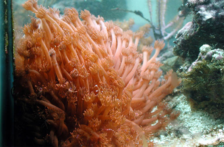
The Goniopora spp. in the author’s tank is an unusual long-term survivor, having been acquired from another aquarist’s tank where it had survived for several years. However, the genus is known to produce strong allelopathic substances that can negatively affect other corals, fish, invertebrates and even aquarists.
Fearon and Cameron (1997) later produced a study where five species of stony corals (_Platygyra daedaelea, Gonaistrea favulus, Favia matthai, Pavona decussata, and Fungia fungites_) were collected, and extracts tested for their effects on the gametes and planulae of G. favulus, P. daedaelea, P. decussata, Oxypora lacera, and_ Pocillopora damicornis_. They were testing to see if extracts were able to inhibit settlement of other coral larvae. They found that the extracts of all five species were lethal to larvae of at least two species at one or more of the concentrations of 62.5, 125, and 250 mg/l. Larvae were found to change their shapes, shrink, cease swimming, and eventually die. Between this study and others (Fearon and Cameron 1996, Koh 1995), larvotoxins produced by stony corals can be summarized below (Table 3). Even though it is now recognized how potently toxic Goniopora tenuidens is, its activity against coral larvae was less than some of the
species studied here.
| Producers | Non-producers |
|---|---|
| Goniopora tenuidens | Porites cylindrica |
| Tubastraea faulkneri | Seriatopora hystrix |
| Platygyra daedaelea | Goniastrea favulus |
| Pavona decussata | |
| Favia matthai | |
| Fungia fungites |
With regard to Goniopora toxin, the chemical is a polypeptide toxin (19,000MW) that is a voltage dependent Ca2+ channel activator, and is highly active at the 5mM level (Qar et al. 1986). It is found in varying amounts in all Goniopora examined to date, and its levels seem to vary temporally and according to external or environmental factors (as is the case with the secondary metabolites of most marine organisms). In the study mentioned above, 95g of Goniopora tissue was sufficient to conduct a large number of tests on a variety or tissues and organisms, including lethal dose assays on mammals. During work for an unpublished thesis by Meredith Peach, she remarked that simply working with Goniopora tenuidens in her studies on feeding ecology produced a reaction on her skin severe enough that she had to wear gloves during contact with the corals or even the water of aquariums housing them (Peach pers. comm.). Many other studies involving the effects of Goniopora toxin on physiological and biochemical processes are listed in the references at the end of this article.
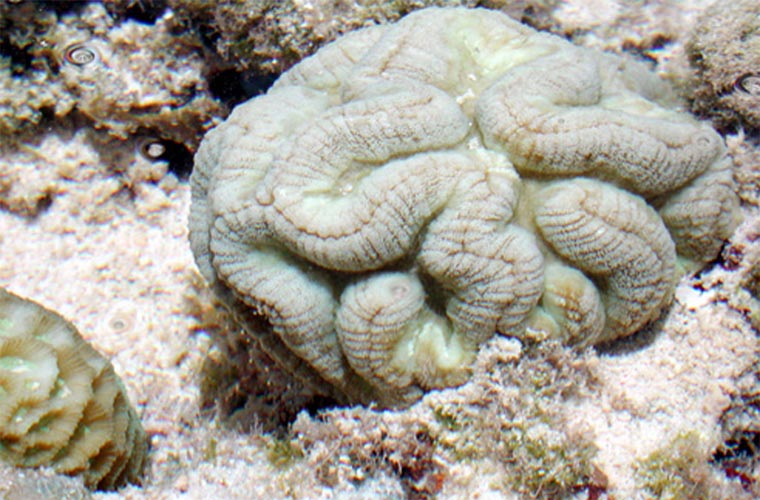
Symphyllia spp. are one of many stony corals that have been found to produce toxic secondary metabolites. Photo: Eric Borneman
At this point, it should be quite apparent that no longer can we assume that toxicity effects in closed systems are only by certain commonly maintained organisms like soft corals. The fact that the aqueous extracts of stony corals causes polyp contraction, bleaching and tissue loss in other stony corals within 24 hours, and that merely putting Galaxea in a tank with Goniopora can cause it to die within hours should put us at a new state of awareness and alert. As with soft corals, it may be difficult to say with certainty that coral species x will have a particular effect on coral species y. As with other organisms, variability in both production and effects on other species seems to be the norm. However, and as I have stated frequently, it becomes a matter of risk assessment. Soft corals known to be prolific producers of toxins, or those known to have certain effects on certain species, should be maintained in aquariums with careful consideration of the potential consequences. It appears the same is true for stony corals. In particular, Goniopora is well recognized to produce toxins that are wide-ranging and consistent in their effects on vertebrates and invertebrates. Perhaps this is fortunate news, given the fact that they survive so poorly in aquariums (Borneman 1997, Toonen 2001). Perhaps our search for the methods to keep these corals alive should be ameliorated or ended in light of their obvious toxic potency. Even more, the continued death of these corals in tanks will certainly result in the release of the total amount of toxins in the tissue throughout the tank. Given the fact that a single colony possesses enough toxins to kill hundreds of mice, this is no small matter of concern for those who purchase these animals in unlikely hopes of having such beautiful corals survive in their tank. Equally as disturbing are the number of other stony corals that may have similar, equal, or even more potent toxicity across various assays. Not that this is really any great surprise to me, although seeing it all spelled out in the aforementioned studies was impressive. I noted in numerous places in Aquarium Corals (2001) where I had observed what seemed to be allelopathic effects by stony corals in my own aquariums – notable Echinophyllia, Oxypora, and Pachyseris species. Similarly, reports of aquarists over the years would lend further support of such effects in closed systems.
For many years, noted authorities have suggested that keeping aquariums with lots and lots of miniature species, eventually bound upon survival to compete with each other in a small closed volume of water, was asking for trouble. Now, more than ever, the effects of such allelopathic competition, even in tanks that do not house significantly large or numerous soft corals, is a very likely occurrence for which a contingency plan should be devised prior to the purchase of numerous, and especially highly toxic, species of corals. I would also suggest that the use of activated carbon maybe among the more effective ways of dealing with such secondary compounds, as it has been found to be effective in the absorption of similar compounds from terrestrial plants. Studies should also be undertaken to determine the composition of protein skimmate to see how efficient these devices are at affecting water concentrations of the organic chemical soup that characterizes out indoor reefs.
Addendum
Terry Siegel kindly asked me to expound on some of the material in the last two paragraphs of this article. He notes quite correctly in an email to me that, “There are many reef keepers, myself included, that have very dense coral growth, and corals that I’ve had for more than 15 years. Why are we able to get away with this?” Before proceeding, I would also note that Siegel also wrote several excellent articles not too long ago about “old tank syndrome,” and has communicated to me numerous times about losses of corals. See: http://www.animalnetwork.com/fish2/aqfm/1999/jan/outer/default.asp and http://www.animalnetwork.com/fish2/aqfm/1998/dec/outer/default.asp
Like him, I think most of us with long-term corals in our tanks might wonder about allelopathic effects, and if they are significant. I would add that, while many of us may have dense coral growth and may have many relatively “old” specimens in our tanks, I feel safe saying this same period has been punctuated by many losses, some of them without obvious explanation. What follows is largely speculative on my part in terms of aquarium topics, but also based on a fairly thorough review of literature on natural products, wastewater treatment, and studies of allelopathy in both terrestrial and aquatic environments.
With that, I would like to further explain the nature of many of these metabolites across taxa, not solely those of stony corals. A single species may have from a few to over a hundred chemicals it produces, some or many of which may have intentional or incidental allelopathic actions. In some studies, there is always damage of species x on species y. In some case, damaging effects may also occur on species a and species c, but never on species b. Thus, their action on tank inhabitants may not be predictable, much less known for certain as relatively few of these compounds have been tested extensively for their action on all but a handful of other species. It is not a requirement that the producer has effects on related species, either. By example, a coral metabolite might have an effect on echinoderms, but has never been reported. By and large, compounds produced by organisms to have an allelopathic role tend to be geared towards organisms that compete with the producer, and this means that related species are often the target – but not always.
Furthermore, in the scope of marine organisms, very few compounds have even been isolated. Because a species is known to produce a compound does not mean it doesn’t produce a dozen others that have not been isolated or identified. Not all chemicals produces are released, and some are stored in the tissues of organisms. In these cases, the toxic effects may not be seen until partial or total mortality of the organism occurs. This is perhaps well illustrated with sexual spawning in _ Caulerpa_ or some corals when spawning results in massive tank mortality. Allelopathy is, for lack of a better phrase, a “grab bag” of chance in most situations.
Compounding these variables are the various environmental factors that affect the production of bioactive compounds. Some are produced seasonally, during reproduction, under stress, under conditions of limiting nutrients, under conditions of abundant resources, when the animal or plant is being grazed, when directly involved in competition, etc. Thus, it is very hard to predict the levels of production of even a species known to produce toxic metabolites. It could very well be that production remains low, and something as relatively simple as a new addition or an injury or a new food causes the animal to ramp up production of various compounds.
In the interest of explaining why tank inhabitants don’t keel over on a regular basis from allelopathic organisms present in aquariums, it may be that environmental conditions, including our deliberate avoidance of predators of specimens we keep in tanks, or the stability of some systems, limits their production. Also likely is a habituation response, whereby either a tolerance to various compounds develops among the other inhabitants, or where the producer habituates to the presence of its co-inhabitants and no longer senses them as an immediate “threat.” I am postulating here, for I do not know for certain what happens in all the potential interactions, but am basing these thoughts on likely scenarios that also occur in nature.
It may also be possible that we are doing an adequate job of removing secondary metabolites from the water. Many of the more toxic compounds studied across terrestrial and marine systems occur in the polar aqueous fractions of extracted tissues. This is not to say that nonpolar compounds with deleterious effects do not exist, but that the majority seems to be polar. As such, they may be more likely to be removed by foam fractionation. In reading literature dealing with allelopathy in both terrestrial and marine systems, as well as copious literature and material from the wastewater industry, it appears that a number of media sources can be employed to remove secondary metabolites. The most commonly employed in scientific methods seems to be activated carbon. While wastewater industry uses activated carbon, they also employ activated clays such as bentonite. Papers in the natural products literature tend to use more sophisticated devices, but the equivalent of deionization cartridges may be useful. In other words, it might work to pump water through various resins if they could be designed to benefit tanks. I am also aware of polymers that selectively absorb compounds, and the aquarium product PolyFilter relies on this technology. The polymers are not specifically designed primarily for these chemicals, but rather those more commonly associated with tank water chemistry issues. However, filters for classes of chemicals could probably be designed and tests of PolyFilters seem to indicate that absorption of similar organics is possible by the products. Obviously, water changes would also be effective in removing levels of metabolites in proportion to the volume of water exchanged. This is probably the simplest, easiest and perhaps most effective way of dealing with such bioactive substances.
There is no reason to suspect that allelopathy, and the simultaneous production of many other compounds that may have other effects, is not occurring in our tanks. Various effects may result, from reactions by other organisms that range from acute toxicity, to a general “failure to thrive,” to no visible effects (even though there may be very significant effects that are simply not visible to the aquarist, such as changes in respiration or photosynthesis rates). There may also be cumulative effects, with low levels produced increasing over time so that levels that initially had no effects begin being expressed over time on various organisms, perhaps in various ways. Such a progressive increase of metabolite concentrations could help explain the “old tank syndrome” Siegel mentioned in his articles, and although there are a host of other potential explanations, the signs are consistent with what one would expect from allelopathy. Finally, allelopathy may likely be a part of the
reason for relatively low levels of sexual reproduction occurring in our aquariums, especially among corals. Once again, I have no reason to suspect that this is THE reason, but studies would suggest it is at least possible if not probable. The number of observations by the reef aquarium community provides substantial anecdotal evidence of such events occurring, but unfortunately only become noticed when conditions in the tank are quite dramatically affected. It is my purpose here not to create widespread panic, or complacency, but to make the aquarium community aware of the widespread occurrence of marine organisms producing scores of bioactive compounds, and to briefly describe the possible effects they produce. In summary, it is my belief that our aquariums are under potentially significant influence by such compounds, and further examination of the effectiveness of methods that mitigate their effects should be a priority.
References
- Ashida K, Toda H, Fujiwara M, Sakiyama F (1987) Amino acid sequence of Goniopora toxin. Jap J Pharmacol 43 Suppl: 187 pp.
- Ashida K, Sakakibara Y, Muramatsu I, Fujiwara M (1983) Molecular structure and vascular relaxing effects of new terpenoid from marine coral. Jap J Pharmacol 32 Suppl: 183 pp.
- Bakus GJ, Targett Nm, Schulte B (1986) Chemical ecology of marine organisms: a review. J Chem Ecol 12: 951-987.
- Borneman EH (1997) A Death In the Family? The Mystery of Goniopora. Aquarium Net November issue. www.aquarium.net.
- Coll, JC, Sammarco PW (1986) Soft corals: chemistry and ecology. Oceanus 29: 33-37.
- De Ruyter van Steveninck ED, Van Mulekom LL, Breeman AM (1988) Growth inhibition of Lobophora variegata (Lamaroux) Womersly by scleractinian corals. J Exp Mar Biol Ecol 115: 169-78.
- Endean R, Cameron AM (1983) Toxins in coral reef organisms. Toxicon suppl 3: 105-109
- Fujiwara M, Hong S-C, Muramatsu I (1982) Effects of Goniopora toxin on non- adrenergic, non-cholinergic response and purine nucleotide release in guinea- pig taenia coli. J Physiol 326: 515-526.
- Fujiwara M, Muramatsu I, Hidaka H, Ikushima S, Ashida K (1979) Effects of Goniopora toxin, a polypeptide isolated from coral, on electromechanical properties of rabbit myocardium. J Pharmcol Exp Ther 210: 153-157.
- Gonoi Y, Ashida K, Feller D, Schmidt J, Fujiwara M, Catterall WA (1986) Mechanism of action of a polypeptide neurotoxin from the coral Goniopora on sodium channels in mouse neuroblastoma cells. Mol Pharm 29: 347-354.
- Grozinger K, Freter K, Farina r, Gladczuk A (1983) Synthesis of 7- and 9-substituted l-methylisoguanines from 4(5)-amino-5(4)-cyanoimidazole. Eur J Med Chem – Chim Ther 18: 221-226.
- Gunthorpe L, Cameron AM (1990a) Toxic exudate from the hard coral Goniopora tenuidens. Toxicon 28(11): 1347-1350.
- Gunthorpe L, Cameron AM (1990b) Widespread but variable toxicity in scleractinian corals. Toxicon 28(10): 199-1219.
- Gunthorpe L, Cameron AM (1990c) Intracolonial variation in toxicity in scleractinian corals. Toxicon 28(10): 1221-1227.
- Hashimoto Y, Ashida K (1973) Screening of toxic corals and isolation of a toxic polypeptide from Goniopora spp. Proc 2nd Int Symp Cnidaria. Publ Seto Mar Biol Lab 20: 703-711.
- Ikushima S, Muramatsu I, Fujiwara M, Ashida K (1981) Relationship between the effects of Goniopora toxin on action potential and on contractile force in guinea-pig papillary muscle. Jap J Pharmacol 31: 1051-1060.
- Ikushima S, Muramatsu I, Fujiwara M (1982) Nicotine-induced response in guinea-pig aorta enhanced by Goniopora toxin. J Pharm Exp Ther 223: 790-794.
- Kaul PN, Kulkarni SK, Wienheimer AJ, Schmitz FJ, Karns TKB (1977) Pharmacologically active substances from the sea. II. Various cardiovascular activities found in the extracts of marine organisms. Lloydia 40: 253-268.
- Koh EGL (1995) Chemical warfare among scleractinians: bioactive natural products from Tubastraea faulkneri Wells kill larvae of potential competitors. J Exp Mar Biol Ecol 251:141-160
- Koh EGL (1997a) Do scleractinian corals engage in chemical warfare against microbes? J Chem Ecol 23: 379-98.
- Koh EGL (1997b) Secretion of bioactive compounds by a scleractinian coral. Proc 8th Int Coral Reef Sym 2: 1263-6.
- Fearon RJ, Cameron AM (1997) Preliminary evidence supporting the ability of hermatypic corals to affect adversely larvae and early settlement stages of hard coral competitors. J Chem Ecol 23(7): 1769-1780.
- Mc Caffery EJ, Endean R (1985) Antimicrobial activity of tropical and subtropical sponges. Mar Biol 89: 1-8.
- Muramatsu I, Fiujiwara M, Miura A, Narahashi T (1985) Effects of Goniopora toxin on crayfish giant axons. J Pharm Exp Ther 234: 307-315.
- Muramatsu I, Fiujiwara M, Ikushima S, Ashida K (1980) Effects of Goniopora toxin on guinea-pig blood vessels. Naunyn-Schmie Arch Pharmacol 312: 193-197
- Noda M, Muramatsu I, Fujiwara M, Ashida K (1985) Effects of Goniopora toxin on bullfrog atrial muscle are frequency dependent. Naunyn-Schmie Arch Pharmacol 330: 59-66.
- Noda M, Muramatsu I, Fujiwara M (1984) Effects of Goniopora toxin on the membrane currents of bullfrog atrial muscle. Naunyn-Schmie Arch Pharmacol 75-80.
- Qar J, Schweitz H, Schmid A, Lazdunski M (1986) A polypeptide toxin from the coral Goniopora: purification and action on Ca2+ channels. FEBS 202(2): 331-336.
- Sammarco PW, Coll JC, LaBarre S (1985) Competitive strategies of soft corals (Coelenterata: Octocorallia). II. Variable defensive responses and susceptibility to scleractinian corals. J Exp Mar Biol Ecol 91: 199-215.
- Sammarco PW, Coll JC (1990) Lack of predictability in terpenoid function: multiple roles and integration with related adaptations in soft corals. J Chem Ecol 16: 273-89.
- Sammarco PW, Coll JC (1987) The chemical ecology of Alcyonarian corals. In: Bioorganic Marine Chemistry Vol. 2, (Paul J. Scheuer, ed.). Springer-Verlag, Berlin: 87-116.
- Sammarco PW, Coll JC, LaBarre S, Willis B (1983) Competitive strategies of soft corals (Coelenterata: Octocorallia): allelopathic effects on selected scleractinian corals. Coral Reefs 2: 173-8.
- Sheppard CRC (1979) Interspecific aggression between reef corals with reference to their distribution. Mar Ecol Prog Ser 1: 237-47.
- Toonen, Rob J. 2001. Goniopora. FAMA 24(6): 142-158

0 Comments