UW professor is digitizing every fish species in the world
by Michelle Ma

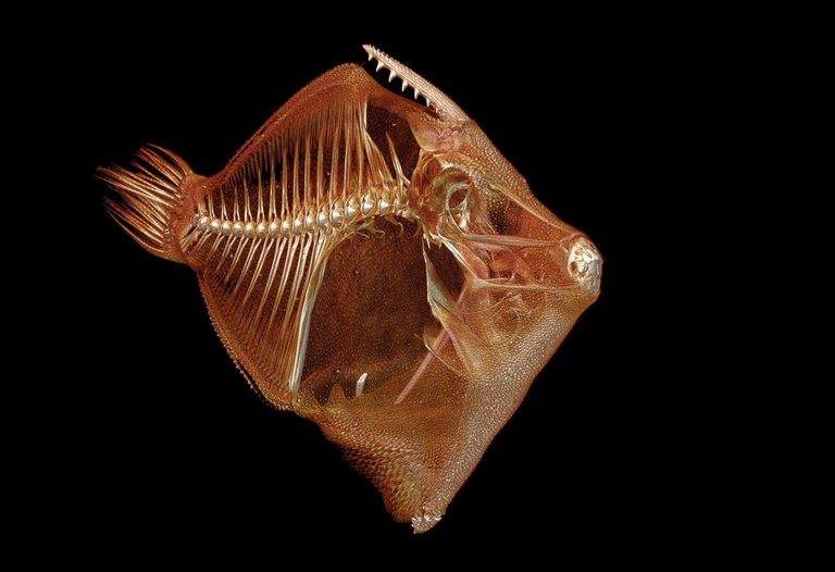
A scan of the spotfin hatchetfish (Thoracocorax stellatus).University of Washington
Nearly 25,000 species of fish live on our planet, and a University of Washington professor wants to scan and digitize them all.
That means each species will soon have a high-resolution, 3-D visual replica online, available to all and downloadable for free. Scientists, teachers, students and amateur ichthyologists will be able to look at the fine details of a smoothhead sculpin’s skeleton, or 3-D print an exact replica of an Arctic alligatorfish.
“These scans are transforming the way we think about 3-D data and accessibility,” said Adam Summers, a UW professor of biology and aquatic and fishery sciences who is spearheading the project.
Summers, who is based at the UW’s Friday Harbor Laboratories, uses a small computerized tomography (CT) scanner in the back room of a lab to churn out dozens of fish scans from specimens gathered around the world. The machine works like a standard CT scanner used in hospitals: A series of X-ray images is taken from different angles, then combined using computer processing to create three-dimensional images of the skeleton.
The goal is to make it possible for scientists to examine the morphology of a particular species, or try to understand why a group of fish all have similar physical characteristics such as bony head “armor” or the ability to burrow into the sand.

Red-throat Piraha (Serrasalmus medinai)
“It’s been so fun to throw this data up on the web and have people actually use it,” Summers said.
Until now, there hasn’t been an easy — or cheap — way for scientists to get detailed, three-dimensional scans of critters. Summers recalls having to beg hospitals to scan his specimens, including a sting ray that in 2000 was the first CT-scanned fish to appear on the cover of a biology journal.
Over the years, Summers and colleagues have developed more efficient ways to scan specimens in larger batches at hospitals, but each scan was still very expensive — anywhere from $500 to $2,000 each.
Summers saw the need for an in-house scanner and raised $340,000 to purchase the machine last November that now lives at Friday Harbor Laboratories. His policy is that it’s free and open to anyone who wants to use it, and the fish must come from museum-accessioned collections.
Students, postdoctoral researchers and professors from around the world have taken Summers up on the offer and come to the labs on San Juan Island to scan their favorite specimens. They also send boxes of fish specimens in the mail for Summers’ lab to scan and post online. Fish from museum collections are trackable with numbers, and the online database now houses scans of fish from the UW’s Burke Museum of Natural History and Culture, the National Academy of Sciences in Philadelphia, Ohio State University, Western Australian Museum and many others.
“We just make it a group effort to get everything done,” Summers said. “Having this scanner has made it clear to me the incredible power of this system if you think about it the right way.”
Most scientists using the 3-D fish data appear to be interested in measuring morphology — the length of a particular bone, for example — or looking for an aspect of anatomy never seen before. The scans allow you to zoom in digitally at many different angles, or print life-sized or larger plastic replicas of a fish.
Summers has fine-tuned and expedited the process by scanning multiple fish at once. He first packs and rolls the specimens together like a burrito in a cylinder that is then placed directly in the scanner. After the machine makes one scan, Summers then digitally separates each 3-D fish into its own file.
Additionally, he doesn’t scan at the highest-possible resolution, because few scientists actually need that much detailed data. This saves more time and digital space — and makes it possible for people to access the files more easily online.
“The way transformative ideas do, these just instantly changed the way we think about scanning specimens,” Summers said. “We went from, ‘Is this possible?’ to scanning whole series of fishes quickly.”
So far about 515 species have been scanned and many are posted online to Open Science Framework, an open-source, sharing website for scholarly projects. Summers expects to finish scanning all of the fish species in the world in two-and-a-half to three years.
His next project is to scan all 50,000 vertebrate species on Earth — an undertaking he thinks is possible within a few years using half a dozen CT scanners like the one at Friday Harbor Laboratories. Colleagues at museums around the U.S. are writing a grant proposal to accomplish this feat.
###
For more information, contact Summers at [email protected]. High-resolution scan images are available for download on Open Science Framework.
[via University of Washington]


0 Comments