
The reef aquarium displays a world of colourful corals and fishes. Among the corals are other invertebrates, many of which rival the corals in color and beauty. The beginning enthusiast typically enjoys the beautiful vistas of reef aquariums, and tries to obtain as many species of coral as possible. The more experienced aquarists often move on to challenges of a more difficult nature, studying in great detail the less visible organisms within their captive reef.
On display, but still not easily seen, is a hidden world of organisms found in every reef tank: the microscopic world of algae and animals that is a key factor in the biological “balance” of the aquarium. This world is rarely focused upon and remains unknown to most enthusiasts. I would like to reveal some of its secrets here.
Equipment
The resolving power of the human eye is about 0.1 mm, which is equal to about the size of the dots that make up the image in a newspaper photograph. This is not nearly sufficient to resolve micro-organisms, let alone to reveal their structural details. We need help! In principle there are two types of tools that you can use to view the “hidden world”: compound microscopes or binocular lens and stereo dissecting scopes. The compound microscope uses transmitted light to enable us to look through objects while the binocular lens and stereo dissecting scopes often use reflected light to look onto objects. This means that a compound microscope is best used for transparent objects, such as a drop of water. The binocular lens and dissecting scopes are perfectly suited for viewing the surface of live rocks or looking in between the grains of the substratum, revealing the microscopic life here. The choice of an appropriate optical system is crucial. If you try to do it the opposite way and look at the surface of a piece of live rock with a microscope or at the life in a drop of water with a binocular lens, you will not see much (if anything at all).
Also important is the way the object is illuminated. Compound microscopes have a configuration of lenses that enlarge the object up to 1000 times and other lenses that condense light through the object. More advanced microscopes have possibilities for using polarised light and phase contrast, which can reveal details in transparent specimens that would otherwise be obscure. Binocular lens and stereo dissecting scopes also need high quality illumination in order to work properly. Remember that the surface you want to view is tiny and needs to be brilliantly illuminated in order to reveal its details. Light sources incorporating fibre optics are often used to direct intense light onto the subject.
Compound and binocular microscopes are available in a wide range of optical quality, and needless to say, with increasing optical quality come increasing prices! What do you need? If you simply want to look at the microscopic organisms, you need only equipment of quality similar to that commonly used in primary and secondary schools (under $1000.00 each, based on prices here in Norway). If you want to photograph the organisms and view the smallest algae and perhaps even look at bacteria, then you need better equipment that will cost from $2000.00 upwards. The photomicrographs shown here were taken through an Olympus BH-2 compound microscope, which is a relatively expensive, research-level microscope marketed during the late 1980s and early 1990s. C&A sells a decent personal compound microscope (named “My First Personal Microscope”) through Amazon.com for less than $100.00 that has reasonable optics and serves this purpose quite admirably.
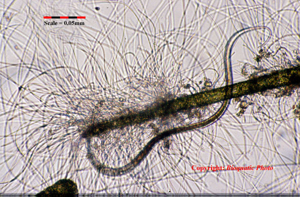
A dramatic scene from a reef tank. A tiny round worm (Phylum Nematoda) is fighting to reveal itself from bacteria and cyanobacteria threads that have grown from the tip of a single thread of filamentous green algae. Magnification on the film plane is 100X. Photo by Bioquatic Photo.
Finding And Logging The Microbes
If you have a microscope or binocular lens available, you still need to find the organisms you want to view. This is sometimes easy, sometimes difficult and sometimes even extremely difficult. It more or less depends on what you are looking for. If you want to look at the delicate details of the filamentous algae growing in your aquarium, this is easy. Scrape some algae from the surface of your aquarium window, place them in a small disk with aquarium water and let them spread out. Pick some threads using a forceps and place them on a glass slide, add one drop of water, cover them with another very thin glass slide and you are ready to go. If you want to look at organisms living in the substrate, you often have to strain the substratum through suitable mesh in order to increase the concentration of the organisms in a small volume of water, and pick your sample from there.
The reef aquarium has macro- and micro-flora and fauna that develop constantly. It is most interesting to log and observe this flora and fauna over time. If one has a microscope and a binocular lens available, this task becomes extraordinary interesting and can really give you valuable information about what goes on inside your aquarium. How do we then go about logging our findings? Luckily, this is quite easily done, although it requires some equipment, patience and work. Apart from the optical equipment you need only:

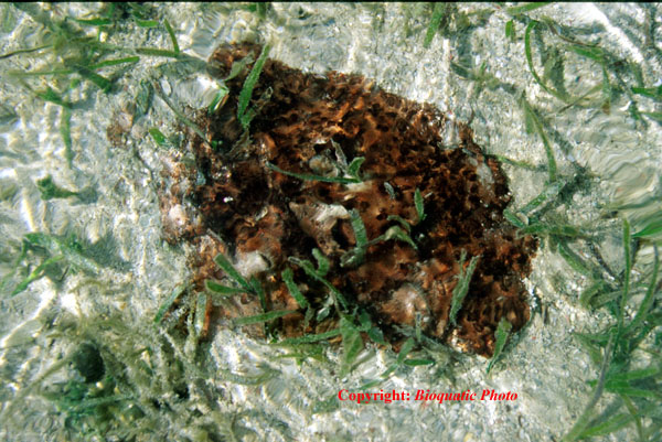
The decomposition of detritus. A detritus particle collected from the back wall of a reef aquarium is colonised with bacteria and Cyanobacteria. 200X magnification on the film plane. Photo by Bioquatic Photo.
- A couple of microscope slides that measure 1 x 3 inches
- A few pieces of floating material like Styrofoam plates or their equivalent.
You now cut two 2 cm slots in the Styrofoam, in a right angle to each other and mount the glass slides in the slots. Be sure to mark the slots so you can identify the individual slides if you use more than one set of slides. Place the mounted glass slides floating in suitable places in your system. If you have access to a digital balance, be sure to weigh the glass slides before they are mounted and make accurate notes on their weight. It is of course possible (and probably advisable) to use more than one pair of glass slides in the same tank.
What spots are “suitable”? I would recommend spots that are not heavily illuminated but which have a steady flow of water. If you have a skimmer or other technical equipment placed in a sump below the aquarium, this is an excellent spot. You can also anchor the slides in the main aquarium along the sides where the light is moderate.
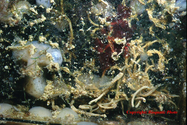
By placing tiny glass sheets inside your aquarium and/or in the sump, you can log the organisms that settle over time. This sheet has been gathering settlers for 16 months in the aquarium itself. Photo by Bioquatic Photo.
What next? You now have to make a plan for examining the slides and logging the organisms regularly. Settlers in your system will very soon start to colonise the slides, which soon will be covered with all sorts of interesting animals. Check the slides at regular intervals, such as on the same day once every two weeks or once a month. By observing the organisms that have settled on the glass slides you get an overview on how many and what types of organisms settle over time, which will be a biological indicator of your captive system. A more advanced logging will be to count the number of different organisms that have settled on fixed areas over time and by using a digital balance (best with an accuracy of +/- 0.1 gram or better) to weigh the slides regularly.
Into The Hidden World
Let us start by looking at some algae-like bacteria. When I started out as a marine enthusiast back in the seventies, the uncontrolled blooming of red filamentous and slimy blue-green “algae” was a huge problem among German reefkeepers. The events were much debated in magazines and clubs. Today, the problem is much less common, although still some aquarists struggle to get rid of the slimy blue-greens. No specific explanation to the explosive growth of the algae-like bacteria has been given. Personally, I believe that the problem had two major causes: an imbalance in nutrient concentrations and more importantly, a far too low salinity. Whatever happened, the blue-green algae are ugly when they take over a reef aquarium, but beautiful when viewed through a microscope.
These Cyanobacteria appeared in a reef aquarium as a black slimy growth on the stem of a soft coral. 400X magnification on the film plane. Photo by Bioquatic Photo.
Today we know approximately 150 genera of blue-green “algae” containing some 2000 species, distributed in wet terrestrial habitats, fresh and saline waters, including the ocean. Blue-green “algae” are also commonly called cyanobacteria, and belong to the super kingdom Prokarya, a group of organisms that may be similar to the first life on Earth, and also includes true bacteria. Today, the cyanobacteria are members of the true bacteria (Eubacteria) and show no taxonomic affiliations to algae at all. The term “blue-green algae” is therefore misleading, accept for the fact that the growth of the cyanobacteria often looks algae-like. (Please refer to http://tolweb.org/tree/phylogeny.html and to http://tolweb.org/tree?group=life#TOC2 for further readings on this subject).
Like all prokaryotes, their cells lack a nucleus. Their geological history dates back more than 3 billion years when colonial stromatolites colonised the ancient shores. Their fossil remains consist primarily of carbonates precipitated because of the locally high pH values induced by photosynthesis. Microfossils corresponding to individual cells can also be found in some stromatolites. Living stromatolites can only be found today in special environments, such as the shallow seas off the coast of Australia where grazing pressure tends to be low. The vivid colours of cyanobacteria are from phycobilin pigments, which often (but not always) block the green Chlorophyll-a, commonly found in nearly all plants.
Cyanobacteria growing among see grass at the Great Barrier Reef. Photo by Bioquatic Photo.
On the reefs blue-green algae are found as patches on sandy bottom, growing as slimy colonies among corals and most importantly as a major component of the overgrowth (or “turf algae”) that cover the rocks and bases of corals. Cyanobacteria are important because they convert relatively inert dinitrogen (N2) into biologically accessible forms and add this nutrient to the ecosystem. This “fixation” of nitrogen takes place in special cells called “heterocysts.” The heterocyst is a large, transparent, thick-walled cell that occurs at intervals along the filament in some blue-green algae. Many blue- green algae consist of long chains of cells that are growing in a polysaccharide matrix (or mucilaginous mass). The matrix envelopes the cells (such as in the genus Anacystis) or in some instances covers the cells in filaments and look almost tube-like (such as in Lyngbya).
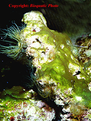
Green forms of Cyanobacteria overgrowing live rock in a reef aquarium. Photo by Bioquatic Photo.
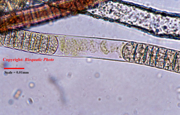
Chain of Cyanobacteria, probably Lyngbya sp. The polysaccharide matrix is clearly visible. 400X on the film plane. Photo by Bioquatic Photo.
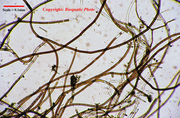
Cyanobacteria (probably Lyngbya sp.) at 40X magnification on the film plane. Photo by Bioquatic Photo.
Now, put some cyanobacteria from your aquarium under a powerful microscope and you might be able to spot these structures and discover the beauty of the blue-greens. The chains of cells are easily caught. With high magnification, heterocysts can be studied and the mucous coating is usually not difficult to see. You will discover that the slimy dark-coloured mass of algae is in fact composed of tiny, beautiful and diversely coloured individual cells.
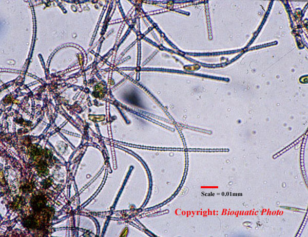
Photo by Bioquatic Photo.
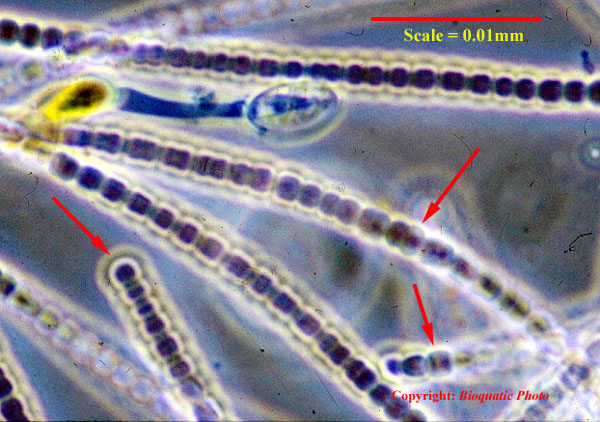
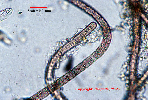
This growth of Cyanobacteria looked like a deep red slimy mass when it was collected from the back wall of a reef aquarium. In the microscope at 200X magnification (film plane) the individual chain of algae are clearly visible. When the magnification is increased to 1000X structures that probably are heterocysts can be seen (arrows). Photo by Bioquatic Photo.
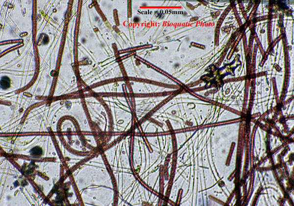
Photo by Bioquatic Photo.

Red forms of Cyanobacteria, commonly known in Germany as “die rote Plage” which have driven aquarists in the early days almost to insanity, look like this when viewed through a microscope at 100X magnification. In AS1240 the magnification is raised to 400X on the film plane. Photo by Bioquatic Photo.
My very first marine aquarium set up in our home here at the island of Hidra in southern Norway became completely overgrown by chocolate brown filming algae shortly after the tank was filled with water. This was caused not by blue-green algae but by diatoms. Diatoms are single celled algae that – besides utilizing the normal algae nutrients – deposit silicic acid in their cell walls. Silicic acid (SiO2nH2O) is commonly found dissolved in ground water. If the concentration of silicon increases above the normal amount found in sea-water (~3000 µg/L), the number of diatoms can explode. This is just what happened in my first marine aquarium. The evaporated water was refilled with fresh water coming from a well, and the water was very rich in silicic acid. As soon as the evaporation was replaced with distilled water, the diatoms decreased to normal populations. And here we touch an important subject: diatoms are important algae in the sea, on the coral reefs and in the coral reef aquarium. Just as I am writing this piece of text the very first blooming of diatoms is occurring in the North Sea just outside my office. The water is colored light yellow and if one uses a microscope, one will discover the beautiful cells of many diatoms. Diatoms are also found among the phytoplankton in the waters surrounding tropical coral reefs, and will always occur as epiphytic algae on other algae, on the reef as well as in the coral reef aquarium. They are important as primary producers and play an important role in the food chain of the coral reefs. In the aquarium these tiny algae are important food for micro- and macro organisms as well as for some fishes, such as bristletooths from the genus Ctenochaetus or the orange shoulder Surgeonfish (_Acanthurus olivaceus_).
Typical growth of diatoms on the decoration of a public marine aquarium. The algae colors the decoration brown. This photo is from Gothenburg, Sweden in 1983 – a time that such scenes were typically seen in public displays. Photo by Bioquatic Photo.
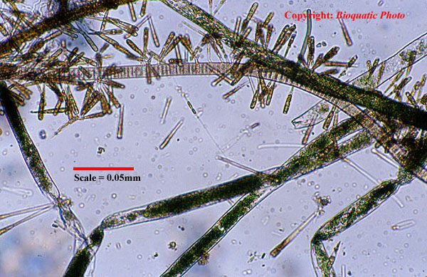
Diatoms growing epiphytic on green thread algae collected in a reef aquarium. This is how diatoms are usually found in our home displays and represent healthy and normal conditions. 100X magnification on the film plane. Photo by Bioquatic Photo.
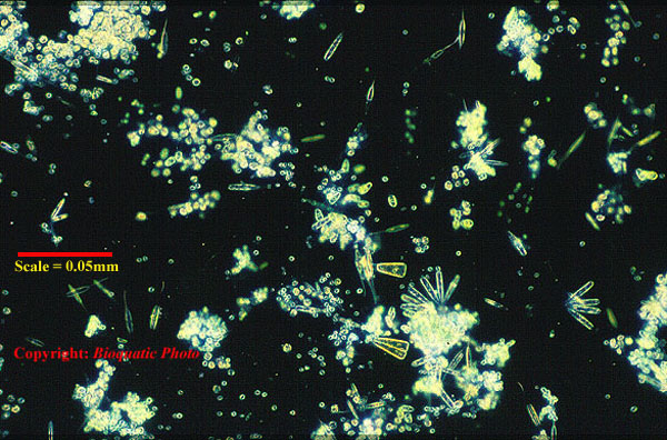
Samples of algae that grew on the front glass of a reef tank viewed under 100X magnification and polarized light (phase contrast) through a microscope. At least three different pinnate diatoms are visible as well as many round flagellates. The dark background is created by manipulating the polarizing light and creates a nice effect that reveals the beauty of the diatoms very well. Photo by Bioquatic Photo.

Three different diatoms and many small, free-living dinoflagellates viewed under 400X magnification. The algae sample was collected from the front glass of a coral reef aquarium. Photo by Bioquatic Photo.
Diatoms have traditionally been placed in “the class Bacillariophyceae,” but are now usually recognised as members of the Stramenopile’s clade, which contains several groups of organisms. As the relationships among these groups are not resolved, the name “Bacillariophyceae” is still commonly used, but expect this to change in the future as more data becomes available.
Two species of epiphytic diatoms growing on green thread algae. We can also spot a free-living diatom. 100X magnification on the film plane. Photo by Bioquatic Photo.
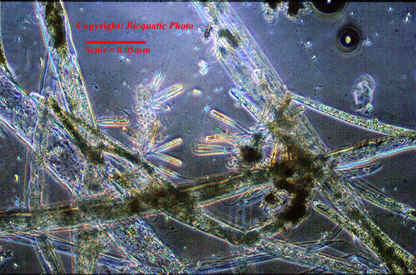
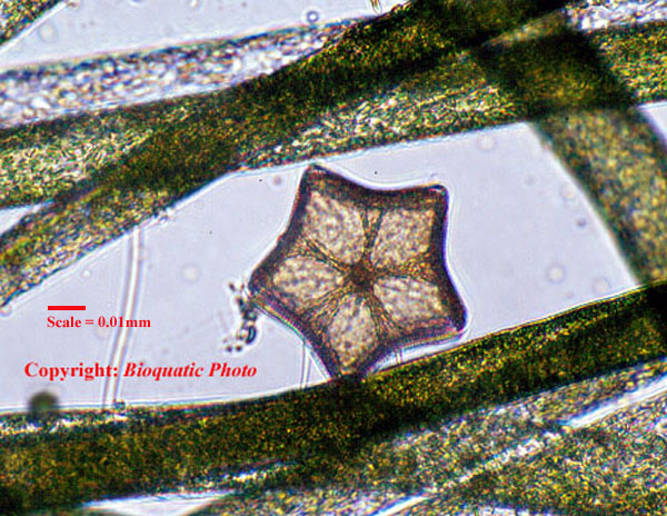
A centric diatom, possibly Bellerochea sp., at 200X on the film plane. We can also see small, pinnate diatoms probably members of the genus Nitzschia. The sample was collected from a reef aquarium. Photo by Bioquatic Photo.
Although there are more than 6000 known species of diatoms, diatoms often consist of an upper and a bottom shell that fit together like the lid and the bottom of a box. (The Latin root of the word diatom “diatoma” means “to cut in half”.) When these algae reproduce asexually the original cell splits lengthwise parallel to the two shells. The resulting two halves become the lids of the new daughter cells, and the bottom half has to be reproduced. Thus, one half of the individuals are reduced in size from generation to generation. When these cells have been reduced to 30 to 40% of their maximum size, they start reproducing sexually, and produce a new generation with maximum size. Given appropriate conditions and sufficient nutrients, the propagation potential is enormous.
A beautiful centric diatom, possibly Biddulphia sp., among green algae in a reef tank. 200X magnification on the film plane. Photo by Bioquatic Photo.
Can we observe the population of diatoms in our reef aquarium? Yes, indeed we can, but as the cells are tiny you have to use a microscope to see the individual cells. The cells are beautiful and often show a number of structures clearly, like the nucleus, chloroplasts, and cell wall. Most diatoms look brown because the brown pigment fucoxanthin masks the green chlorophyll. The class is divided in two orders: pennate and centric diatoms, which is based on the composition of the cell wall. In pinnate diatoms, the cell is elongated and has a bilateral symmetry, whereas centric diatoms have radial symmetry. The cell walls of marine diatoms are highly ornamented and beautiful to the human eye. The intricate design is produced by arrangements of tiny holes in the cell wall through which gas, water, and nutrients are exchanged.
I have found it best to collect diatoms from a reef aquarium by letting the algae settling on the front glass grow for some days and then carefully scrapping off the growth with a razor blade. I collect several scrapes in a small container with a little aquarium water and suck drops of the content with a pipette. The content is placed on the glass slide and placed carefully under the microscope. The pictures following this section tell the story of what is commonly found.
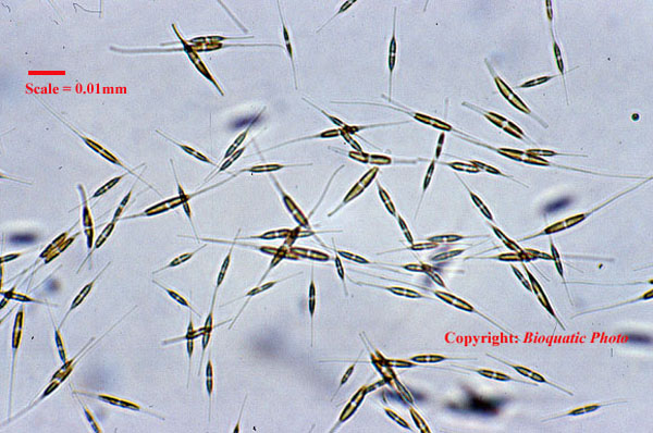
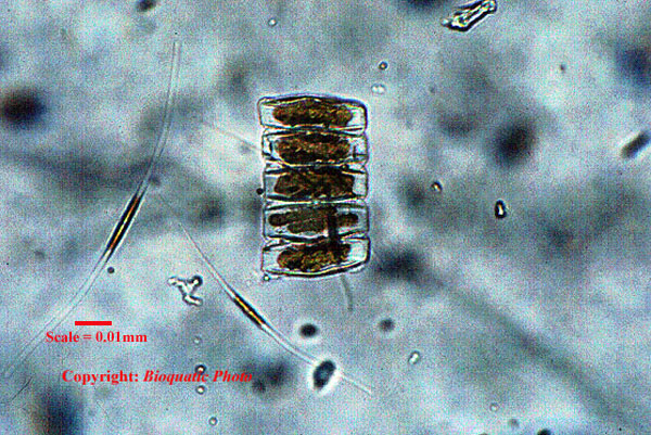
Several small, pinnate diatoms probably members of the genus Nitzschia, as seen through a microscope at 200X magnification. Photo by Bioquatic Photo.
Diatoms and blue-green algae are only two of many groups of organisms that can be studied using a microscope. In the next portion of this article series we shall take a closer look at reproduction among a couple of other algae groups. To be continued…

impressive work you have done.