

A long time ago, back in 1978 at my friend’s room I saw my very first marine aquarium. I did not realise it just then, but my friend’s aquarium was very different from what was marine set ups those days. Instead, of the then, commonly used graveyard-white coral skeletons, calcareous rocks were used as decoration. Instead of changing the water regularly and cleaning the dead coral of all sorts of algae, green thread algae were allowed to grow and flourish. The tank appeared green and exciting as the long green threads of algae swung back and forth. But, most importantly, the fishes recovered completely from a parasite infection that they had when bought in the local pet shop.
In this aquarium the green thread algae represented something new, biologically as well as aesthetically. For the first time we discovered that some sort of “biological balance” could be achieved in a closed aquarium system, and that it was indeed possible to cure disease and prevent the fishes from being further attacked by parasites. The solution was, however, not copper sulphate but the growth of algae!
To-day the “green aquarium,” overgrown by thread algae, is not loved by many aquarists. Although the algae have a positive influence on the well being of many fishes, it tends to overgrow corals and other sessile invertebrates. Let us for a moment forget about this, and observe the general beauty and the interesting reproductive biology of one of the algae that appear as long green threads in the marine aquarium. We are referring to members of the genus Derbesia.
The many species of Derbesia are members of green algae (division Chlorophyta), where they belong to order Caulerpales, in the family Derbeciaceae. The genus Derbesia seems to be cosmopolitic with species found in cold, temperate and tropical waters. The most commonly mentioned species is Derbesia marina, which is reported from Polynesia (Payri & al., 2000), from the Caribbean (Littler & al, 1989), from the Mediterranean (Riedl, 1983) and from cold waters, such as from the Atlantic, North-Sea, and along the Norwegian coast (Nicholls, 1976). I have observed the algae myself in the Maldives and along the coast of Norway, and as mentioned above, is found it to be very common and often abundant in marine aquariums.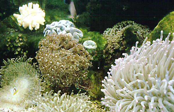
Before we continue, it is necessary to address the reproductive life history of algae in general:
The life history of all plants follows a single basic pattern where there is an alteration between a haploid, gamete-producing phase (the gametophyte) and a diploid, spore-producing phase (the sporophyte). In most plants one of the two is non-photosynthetic and cannot grow independently from the other. In some groups – such as the ferns and many algae – the two phases are so distinct, and independent of each other that the connection between them would not be suspected if the fate of the reproductive bodies that they each produce was not known. Very simplified the life history among algae can be separated into three groups:
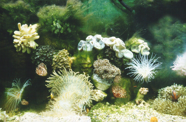
Figure 1 + 2: My first marine aquarium from 1979 was covered with green thread algae. The invertebrates, which at that time were rarely seen in the local pet shops, were soon overgrown by the algae, but the fishes remained healthy and recovered from parasite attacks and infection they had when bought.
- Those with a single morphological phase producing haploid or diploid cells that develop into new plants.
- Those with two morphological phases where a haploid gametophyte produces gametes that fuses and grows to a sporophyte, which in turn produces diploid spores.
- Several red algae with three morphological phases involving a haploid gametophyte, a diploid carposporophyte developing on female gametophyte, and an independent diploid tetrasporophyte.
Derbesia spp fall within group 2, while the red algae of the genus Ceramium – commonly found in marine aquariums and which we deal with later on in this article – belong to group 3. (Table 1 gives some details about which genera of algae that belong in the three different groups).
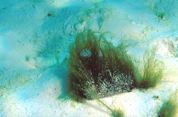
Figure 3: Growth of Derbesia in the shallow water, close to the beach of a coral cay in the Maldives, at about two metres in depth.
Algae reproduction terminology:
- Haploid
- A cell, nucleus, or organism, having a single set of unpaired chromosomes.
- Diploid
- A cell, nucleus, or organism having two haploid sets of chromosomes; two copies of each chromosomes are present, except (in some cases) for the sex chromosomes.
- Diplohaplont
- An organism characterized by alternating diploid and haploid generations.
- Sporophyte
- Plants with alternation of generations, the diploid plant that produces haploid spores.

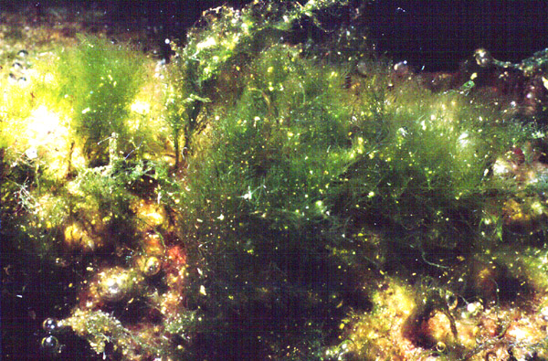
Figure 4: Growth of Derbesia from live rock in a reef aquarium.
- Gametophyte
- An individual plant, or a haploid generation of a plant exhibiting alternating generations, that produces gametes.
- Tetrasporophyte
- A diploid sporophyte (see above) producing spores in groups of four, which are released from a tetrasporangium.
- Carposporophyte
- The diploid sporophyte developing on the female gametophyte, producing haploid or diploid spores.
- Gametangia
- Any cell or organ that produces gametes.
- Isomorphic
- Identical or similar in shape, structure, or appearance.
- Heteromorphic
- An organism that has different forms during different seasons, during different stages of its life cycle, or from generation to generation.
- Spores
- A single-celled or multi-cellular, asexual, reproductive or resting body that is resistant to unfavorable environmental conditions and that is capable of developing into an adult without fusion with another cell when the environment is favorable.
- Zoospores
- Motile spores that can move for instant by the means of flagella.
- Gametes
- A mature haploid reproductive cell that unites with another such cell of the opposite sex to form a diploid zygote. Typically sperm and egg.
- Antheriduim
- Structure on red algae gametophytes that produces male gametes
- Oogonium
- Structure on red algae gametophytes that produces female gametes.
| Group | Haploid | Diploid |
|---|---|---|
| Rhodophyta | Dunaliella | Codium, Caulerpa, Udotea, Halimeda |
| Gametophyte | None | Fucus, Sargassum |
| Others | Dinoflagellates | Diatoms, some dinoflagellates |
| Group | Isomorphic | Heteromorphic | |
|---|---|---|---|
| Gametophyte | Sporophyte | ||
| Chlorophyta | Ulva, Cladophora, Enteromorpha | Monostroma, Acrosiphonia, Halicystis, Bryopsis | Codiolum, Derbesia |
| Phaeophyta | Ectocarpus, Dictyota | Scytosiphon, Petalonia, Laminarian gametophytes | Laminaria |
| Rhodophyta | – | Porphyra, Bangia | Conchocelis |
| Group | Isomorphic | Heteromorphic | |
|---|---|---|---|
| Gametophyte | Tetrasporophyte | ||
| Rhodophyta | Chondrus, Delesseria, Eucheuma, Gracilaria, Griffithsia, Polysiphonia | Bonnemaisonia, Gigartina, Acrosymphyton | Traillella, Petrocelis, Hymenoclonium, Ceramium |
The exciting thing is that although the filamentous growth of Derbesia might look ugly and unpleasant, the life history of the slimy threads is indeed interesting and highly observable in the marine aquarium if you look at the algae threads through a microscope.

Figure 5: The thalli of Derbesia at 100X magnification on the film plane. The chloroplasts are clearly visible, but note that the green colour is lacking from the tip of the individual thallus where the thread grows.
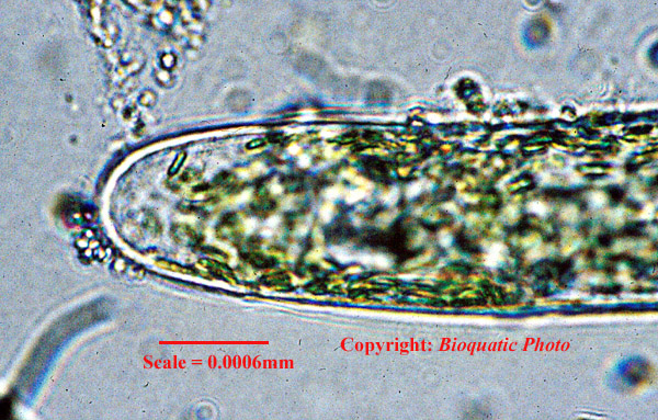
Figure 6: The growth tip of a Derbesia thread at high magnification (1000X on the film plane). The cell wall, the individual chloroplasts and the water filled vesicles are clearly visible.
Put some Derbesia threads under your microscope and use 40 or 100X magnification and you will easily see the thalli (the body of plants that cannot be divided in root, stem or leaves) being composed of delicate and rather beautiful threads that can grow to 10 cm in length. The green colour reflects the chlorophyll pigments that are stored in chloroplasts, and even at this low magnification you can see that the chloroplasts are scattered inside the threads with liquid-filled vesicles (vacuoles) in between. Increase the magnification to 1000X and you will be able to see the individual plastids and the cell wall, which is composted of mannan. Perhaps you can also spot some of the many nuclei?
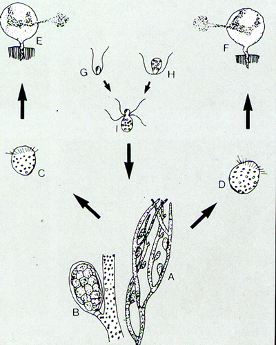
Drawing 1: The reproductive cycle of Derbesia (from Hoek & Al (1978) In Fosså & Nilsen (1996).
Derbesia is a heteromorphic diplohaplont. This means that during a complete life cycle the algae alters between a haploid and a diploid stage that appear differently. The sporophyte is the algae threads (A), which develop pear- shaped sporangia (B) that are attached to the algae threads by a little stalk. Inside the sporangia, zoospores (C & D) develop by the means of meiosis. About half of the zoospores settle and develop to female gametophytes while the other half settle and develop to male gametophytes (E & F). The gametophytes grow to about 5 mm height and are known as Halicystis-stages. The Halicystis contain chloroplasts and many nuclei and a large vacuole in the centre. The fertile male gametophyte develops light-green patches and male gametes are released through pores (E & G). The female gametophyte develops dark-green patches and releases female gametes (F & H). The gamete fuses to a zygote (I) that settles and grows to a new sporophyte. The life cycle is completed.
Continue to view the algae threads at low magnification, and it will not be long until you discover pear-shaped outgrowths on the threads. These are sporangia and give you the first hint that you are approaching the reproductive cycle of the green thread algae. Now it gets exciting! (Please keep your eye on drawing 1 and text outlining the reproductive cycles of this species).
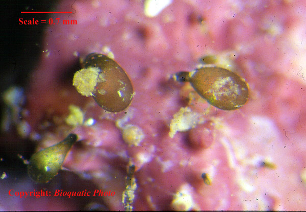
Figure 7: Halicystic stages of Derbesia. Light green gametophytes grow from a live rock in a reef aquarium. 9X magnification on the film plane.
The first time I saw the sporangia myself, I did not realise what this actually was. I did, however, locate three of these structures that grew side by side and placed them in the centre of the microscope’s field of view. It was clear that the three were in different stages of development as the one in the middle was much lighter green that the other two were coloured very dark green. The one to the right in the field of view had also its cell membrane expanded more than the other two. And then, as the strong light from the microscope heated the object viewed, this sporangium exploded releasing all its zoospores in a few seconds.
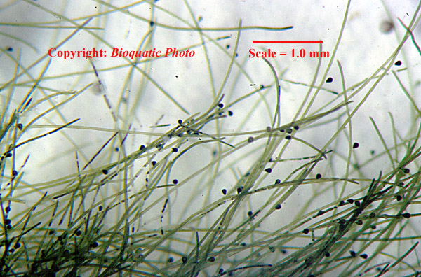
Figure 8: The sporophyte with sporangia as it appears through a binocular lens at 6X magnification on the film plane.
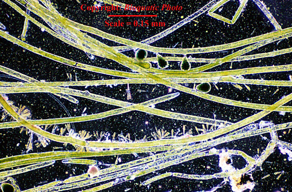
Figure 9: The sporophyte and sporangia as seen through a microscope at 40X magnification on the film plane. Note the many epiphytic diatoms, a free living diatom, and what are probably zoospores and small free-living flagellates scattered all over the field of view.

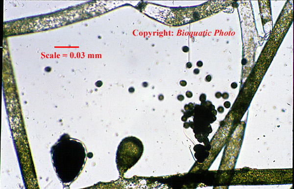
Figure 10 + 11: Three sporangia have grown from the thallus of a Derbesia sporophyte. Note that the middle sporangium is immature and light green while the two others are mature and darker. The right sporangium has the cell membrane expanded. When the heat from the microscope’s lamp warms up the object, the right sporangium explodes releasing the zoospores. 100X on the film plane.
Luckily, I managed to have it all in focus and was there to capture it all on film. It was one of those moments you do not forget. It can very well illustrate how much biology a reef aquarium teach!
The zoospores moved rapidly away with their circulating movements, and as I looked at other samples I discovered that zoospores from Derbesia were everywhere.
When I later on examined the surface of some live rock pieces covered with red calcareous algae, I discovered what I first believed to be ball-shaped algae from the genus Valonia or Cyrtocaria, but later on realised that what I actually found was the gametophytes of Derbesia (known as Halicystis-stages, though resembling Valonia ).
By altering between a sexual and non-sexual stage the green thread algae manage to spread most efficiently and at the same time secure genetic variability. By using a microscope you will be able to observe this interesting piece of biology close up!
A Red Algae
If the life cycle of Derbesia seemed complex, those of red algae (Rhodophyta) are even more complex. As Table 2 shows, many red algae have life cycles that involve three different stages. Among these are members of the genus Ceramium, red alga that typically appear in the marine aquarium.
Ceramium is a diverse genus with cosmopolitan distribution. Most species are turf-like in colder waters most, and are often found from the mid littoral zone and downwards. On the coral reef, some species of Ceramium are members of the turf algae, commonly found on reef boulders and at the base of corals. Payri & al. (2000) mention six species from French Polynesia, while Ceramium rubrum is a common species along the coasts of Europe and is listed in many publications (such as Campbell, 1977). Littler & al. (1989) mention the genus from the Caribbean and Brazil, while Abbott (1997) lists 6 species from Hawaii.
A typical life cycle of red algae is outlined in Drawing-2. In general, a typical life cycle is like this: A diploid tetrasporophyte develop sporangia and produces tetra spores by meiosis. The tetra spores develop to male and female gametophytes, which develop male and female gametangia that produce gametes. The male gametes are produced in structures called antheridia, while female gametes are produced in oogonia, which often develop as a sort of container or cavity on the gametophyte. The male gametes enter the oogonia where the fertilization occurs. The fertilized egg cell develops to a haploid carposporophyte, which is an almost invisible parasitic plant living attached to the mother gametophyte. The carpo-sporophyte does in turn produce carpospores that grows to a new diploid tetrassporophyte.
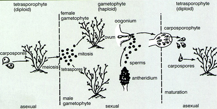
Drawing 2: Outline of a typical red algae life cycle. After Wallace & al. (1986) in Fosså & Nilsen (1996).
Obviously the reproductive cycle in red algae is very complex. Using a microscope I have been able to detect carposporophytes of Ceramium as shown in figures 12 and 13. So far I have not been able to locate sporangia myself, but I encourage aquarists to examine their red algae to possibly reveal more of the red algae’s complex but interesting life cycle.
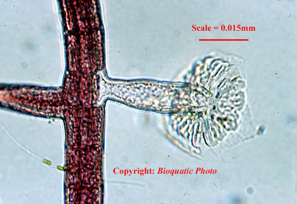
Figure 12: Close up of the carposporophyte on Ceramium. 400X on the film plane. Do also note the shape of the individual cells in the alga thallus.
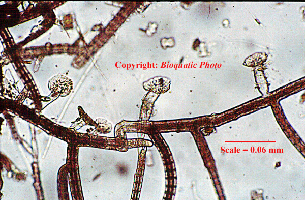
Figure 13: Thalli and Carposporophytes of Ceramium. 100X on the film plane.
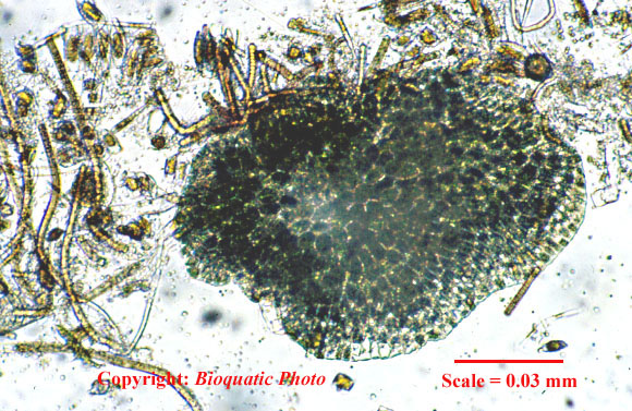
Figure 14: Unknown green alga growing on the front glass of a small reef tank. 200X on the film plane.
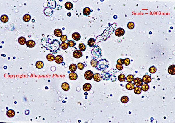
Figure 15: Zooxanthellae in close up. Sample from Euphyllia ancora. 400X magnification on the film plane.
In the next part of this series we shall take a look at some of the peculiar animals that you can find hunting with a microscope in your reef aquarium. However, I cannot let you go just yet…
A few years back, when I operated a small experimental aquarium (Nilsen, 1999), I spotted a strange patchy growth of green algae on the front glass of my tank. I used the microscope and found a strange alga as shown in figure 14. Still this alga remains unidentified, even to genus. Can anyone help identify it?
And at the end: Do not forget that you can use the microscope to take a closer look at the most important of all algae on the coral reef and in the coral reef aquarium – the zooxanthellae. The symbiotic algae belong to the genus Symbiodinium. Traditionally only one single species was believed to exist ( S. microadriaticum ), but to day we know that several species exist and that the algae develop specific strains as a response to environmental differences.
Cut a tentacle of a broad polyp stony coral (such as Euphyllia sp.) or cut a little piece of a soft coral branch. Squeeze the tissue gently and let the tissue fluid of the sample spread on the microscope slide. Cover it and use your microscope. You will easily observe the tiny brown dinoflagellates that are responsible for the growth and ecological success of the tropical giant underwater structure called “the coral reef”.
References
- Abbott, I. (1997): http://www.botany.hawaii.edu/reefalgae/redskey.htm
- Campbell, A.c. (1977) Planter Og Dyr I Grunne Farvann. Gyldendal Norsk Forlag, Oslo, Norway (in Norwegian). original Edition: the Hamlyn Guide To The Seashore And Shallow Seas Of Britain And Europe. The Hamlyn Publishing Group Ltd. (1976), U.k.
- Dring, M. J. 1986. The Biology Of Marine Plants. Edward Arnold, London, U.k. 199 Pp.
- Fosså, S. A. & A. J. Nilsen (1996): the Modern Coral Reef Aquarium Vol. 1.
- Edition. Birgit Schmettkamp Verlag (bsv), Bornheim, Germany.
- Hoek, C. Vand Den 1984 algen. 2. Ausgabe. Thieme Verlag, Stuttgart, Gernany. 481 Pp.
- Lillter, D. S., M. M. Littler, K. E. Bucher And J. N. Norris. (1989). marine Plants Of The Caribbean. Airelife Publishing Ltd., Shrewsbury, U.k.
- Nilsen, A.j. (1999): A Small Experimental Aquarium. marine Fish And Reef 1(1):102-120.
- Payri, C, A De R. N’yeurt And J. Orempuller (2000). algae Of French Polynesia. Au Vent Des Iles, Tahiti (isbn 2-909790-82-7).
- Riedl, R. (ed.) (1983): fauna Und Flora Des Mittelmeeres. Parey Verlag, Hamburg-berlin.
- Wallace, R. A., J. L. King & G. P. Sanders (1986): biology The Science Of Life. 2. Edition. Scott, Foresman And Company, Usa. 1217 Pp.

0 Comments